[English] 日本語
Yorodumi Papers
- Database of articles cited by EMDB/PDB/SASBDB data -

+
Search query

Keywords
Structure methods
Author
Journal
IF

-
Structure paper

TitleSequential membrane- and protein-bound organelles compartmentalize genomes during phage infection.
Journal, issue, pagesCell Host Microbe, Vol. 33, Issue 4, Page 484-497.e6, Year 2025
Publish dateApr 9, 2025
AuthorsEmily G Armbruster / Phoolwanti Rani / Jina Lee / Niklas Klusch / Joshua Hutchings / Lizbeth Y Hoffman / Hannah Buschkaemper / Eray Enustun / Benjamin A Adler / Koe Inlow / Arica R VanderWal / Madelynn Y Hoffman / Daksh Daksh / Ann Aindow / Amar Deep / Zaida K Rodriguez / Chase J Morgan / Majid Ghassemian / Thomas G Laughlin / Emeric Charles / Brady F Cress / David F Savage / Jennifer A Doudna / Kit Pogliano / Kevin D Corbett / Elizabeth Villa / Joe Pogliano /
PubMed AbstractMany eukaryotic viruses require membrane-bound compartments for replication, but no such organelles are known to be formed by prokaryotic viruses. Bacteriophages of the Chimalliviridae family ...Many eukaryotic viruses require membrane-bound compartments for replication, but no such organelles are known to be formed by prokaryotic viruses. Bacteriophages of the Chimalliviridae family sequester their genomes within a phage-generated organelle, the phage nucleus, which is enclosed by a lattice of the viral protein ChmA. We show that inhibiting phage nucleus formation arrests infections at an early stage in which the injected phage genome is enclosed within a membrane-bound early phage infection (EPI) vesicle. Early phage genes are expressed from the EPI vesicle, demonstrating its functionality as a prokaryotic, transcriptionally active, membrane-bound organelle. We also show that the phage nucleus is essential, with genome replication beginning after the injected DNA is transferred from the EPI vesicle to the phage nucleus. Our results show that Chimalliviridae require two sophisticated subcellular compartments of distinct compositions and functions that facilitate successive stages of the viral life cycle.
External linksCell Host Microbe / PubMed:40168997
MethodsEM (subtomogram averaging) / EM (tomography)
Resolution10.0 - 33.68 Å
Structure data

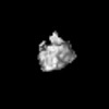
EMDB-48856: 70S Ribosome of Goslar infected WT E. coli
Method: EM (subtomogram averaging) / Resolution: 33.68 Å

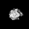
EMDB-48875: 70S Ribosome of Goslar infected chmA KD E. coli
Method: EM (subtomogram averaging) / Resolution: 20.56 Å

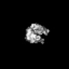
EMDB-48876: 70S Ribosome of Goslar infected chmA KD E. coli
Method: EM (subtomogram averaging) / Resolution: 21.33 Å

EMDB-49120: In situ cryoET of an EPI vesicle in a Goslar infected chmA KD E. coli cell 90 mpi
Method: EM (tomography) / Resolution: 18.0 Å

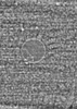
EMDB-49121: In situ cryoET of an EPI vesicle in a Goslar infected chmA KD E. coli cell 90 mpi
Method: EM (tomography) / Resolution: 15.0 Å

EMDB-49122: In situ cryoET of an EPI vesicle in a Goslar infected chmA KD E. coli cell 30 mpi
Method: EM (tomography) / Resolution: 10.0 Å

EMDB-49123: In situ cryoET of an EPI vesicle in a Goslar infected WT E. coli cell 1 mpi
Method: EM (tomography) / Resolution: 10.0 Å

Source
  • Escherichia coli (E. coli)

+
About Yorodumi Papers

-
News

-
Feb 9, 2022. New format data for meta-information of EMDB entries

New format data for meta-information of EMDB entries

  • Version 3 of the EMDB header file is now the official format.
  • The previous official version 1.9 will be removed from the archive.

Related info.:EMDB header

External links:wwPDB to switch to version 3 of the EMDB data model

-
Aug 12, 2020. Covid-19 info

Covid-19 info

URL: https://pdbj.org/emnavi/covid19.php

New page: Covid-19 featured information page in EM Navigator.

Related info.:Covid-19 info / Mar 5, 2020. Novel coronavirus structure data

+
Mar 5, 2020. Novel coronavirus structure data

Novel coronavirus structure data

Related info.:Yorodumi Speices / Aug 12, 2020. Covid-19 info

External links:COVID-19 featured content - PDBj / Molecule of the Month (242):Coronavirus Proteases

+
Jan 31, 2019. EMDB accession codes are about to change! (news from PDBe EMDB page)

EMDB accession codes are about to change! (news from PDBe EMDB page)

  • The allocation of 4 digits for EMDB accession codes will soon come to an end. Whilst these codes will remain in use, new EMDB accession codes will include an additional digit and will expand incrementally as the available range of codes is exhausted. The current 4-digit format prefixed with “EMD-” (i.e. EMD-XXXX) will advance to a 5-digit format (i.e. EMD-XXXXX), and so on. It is currently estimated that the 4-digit codes will be depleted around Spring 2019, at which point the 5-digit format will come into force.
  • The EM Navigator/Yorodumi systems omit the EMD- prefix.

Related info.:Q: What is EMD? / ID/Accession-code notation in Yorodumi/EM Navigator

External links:EMDB Accession Codes are Changing Soon! / Contact to PDBj

+
Jul 12, 2017. Major update of PDB

Major update of PDB

  • wwPDB released updated PDB data conforming to the new PDBx/mmCIF dictionary.
  • This is a major update changing the version number from 4 to 5, and with Remediation, in which all the entries are updated.
  • In this update, many items about electron microscopy experimental information are reorganized (e.g. em_software).
  • Now, EM Navigator and Yorodumi are based on the updated data.

External links:wwPDB Remediation / Enriched Model Files Conforming to OneDep Data Standards Now Available in the PDB FTP Archive

-
Yorodumi Papers

Database of articles cited by EMDB/PDB/SASBDB data

  • Database of articles cited by EMDB, PDB, and SASBDB entries
  • Using PubMed data

Related info.:EMDB / PDB / SASBDB / Yorodumi / EMN Papers / Changes in new EM Navigator and Yorodumi

Read more