Journal: Cell Host Microbe / Year: 2025 Title: Sequential membrane- and protein-bound organelles compartmentalize genomes during phage infection. Authors: Emily G Armbruster / Phoolwanti Rani / Jina Lee / Niklas Klusch / Joshua Hutchings / Lizbeth Y Hoffman / Hannah Buschkaemper / Eray Enustun / Benjamin A Adler / Koe Inlow / Arica R VanderWal ...Authors: Emily G Armbruster / Phoolwanti Rani / Jina Lee / Niklas Klusch / Joshua Hutchings / Lizbeth Y Hoffman / Hannah Buschkaemper / Eray Enustun / Benjamin A Adler / Koe Inlow / Arica R VanderWal / Madelynn Y Hoffman / Daksh Daksh / Ann Aindow / Amar Deep / Zaida K Rodriguez / Chase J Morgan / Majid Ghassemian / Thomas G Laughlin / Emeric Charles / Brady F Cress / David F Savage / Jennifer A Doudna / Kit Pogliano / Kevin D Corbett / Elizabeth Villa / Joe Pogliano / Abstract: Many eukaryotic viruses require membrane-bound compartments for replication, but no such organelles are known to be formed by prokaryotic viruses. Bacteriophages of the Chimalliviridae family ...Many eukaryotic viruses require membrane-bound compartments for replication, but no such organelles are known to be formed by prokaryotic viruses. Bacteriophages of the Chimalliviridae family sequester their genomes within a phage-generated organelle, the phage nucleus, which is enclosed by a lattice of the viral protein ChmA. We show that inhibiting phage nucleus formation arrests infections at an early stage in which the injected phage genome is enclosed within a membrane-bound early phage infection (EPI) vesicle. Early phage genes are expressed from the EPI vesicle, demonstrating its functionality as a prokaryotic, transcriptionally active, membrane-bound organelle. We also show that the phage nucleus is essential, with genome replication beginning after the injected DNA is transferred from the EPI vesicle to the phage nucleus. Our results show that Chimalliviridae require two sophisticated subcellular compartments of distinct compositions and functions that facilitate successive stages of the viral life cycle.
Focused ion beam - Instrument: OTHER / Focused ion beam - Ion: OTHER / Focused ion beam - Voltage: 30 / Focused ion beam - Current: 0.03 / Focused ion beam - Duration: 600 / Focused ion beam - Temperature: 78 K / Focused ion beam - Initial thickness: 200 / Focused ion beam - Final thickness: 140 Focused ion beam - Details: The value given for _em_focused_ion_beam.instrument is Aquilos 2 dual beam microscope. This is not in a list of allowed values {'OTHER', 'DB235'} so OTHER is written into the XML file.
-
Electron microscopy
Microscope
TFS KRIOS
Image recording
Film or detector model: GATAN K3 (6k x 4k) / Average electron dose: 3.8 e/Å2
Electron beam
Acceleration voltage: 300 kV / Electron source: FIELD EMISSION GUN
In the structure databanks used in Yorodumi, some data are registered as the other names, "COVID-19 virus" and "2019-nCoV". Here are the details of the virus and the list of structure data.
Jan 31, 2019. EMDB accession codes are about to change! (news from PDBe EMDB page)
EMDB accession codes are about to change! (news from PDBe EMDB page)
The allocation of 4 digits for EMDB accession codes will soon come to an end. Whilst these codes will remain in use, new EMDB accession codes will include an additional digit and will expand incrementally as the available range of codes is exhausted. The current 4-digit format prefixed with “EMD-” (i.e. EMD-XXXX) will advance to a 5-digit format (i.e. EMD-XXXXX), and so on. It is currently estimated that the 4-digit codes will be depleted around Spring 2019, at which point the 5-digit format will come into force.
The EM Navigator/Yorodumi systems omit the EMD- prefix.
Related info.:Q: What is EMD? / ID/Accession-code notation in Yorodumi/EM Navigator
Yorodumi is a browser for structure data from EMDB, PDB, SASBDB, etc.
This page is also the successor to EM Navigator detail page, and also detail information page/front-end page for Omokage search.
The word "yorodu" (or yorozu) is an old Japanese word meaning "ten thousand". "mi" (miru) is to see.
Related info.:EMDB / PDB / SASBDB / Comparison of 3 databanks / Yorodumi Search / Aug 31, 2016. New EM Navigator & Yorodumi / Yorodumi Papers / Jmol/JSmol / Function and homology information / Changes in new EM Navigator and Yorodumi
 Movie
Movie Controller

 Yorodumi
 Open data
 Basic information
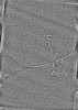
 Map data
 Sample
 Keywords
 Authors
 United States, 1 items
  Citation

 Structure visualization
 Downloads & links
 EMDB map data format
 emd_49121.png
 http://ftp.pdbj.org/pub/emdb/structures/EMD-49121
 Z (Sec.)
 Y (Row.)
 X (Col.)


 Sample components
 Processing
 Electron microscopy
 FIELD EMISSION GUN
