 Movie
Movie Controller
 Structure viewers
 About Yorodumi Papers


+Search query
-Structure paper
| Title | Cryo-EM phase-plate images reveal unexpected levels of apparent specimen damage. |
|---|---|
| Journal, issue, pages | J Struct Biol, Vol. 216, Issue 4, Page 108150, Year 2024 |
| Publish date | Nov 12, 2024 |
 Authors Authors | Jonathan Remis / Petar N Petrov / Jessie T Zhang / Jeremy J Axelrod / Hang Cheng / Shahar Sandhaus / Holger Mueller / Robert M Glaeser /  |
| PubMed Abstract | Apoferritin (apoF) is commonly used as a test specimen in single-particle electron cryo-microscopy (cryo-EM), since it consistently produces density maps that go to 3 Å resolution or higher. When ...Apoferritin (apoF) is commonly used as a test specimen in single-particle electron cryo-microscopy (cryo-EM), since it consistently produces density maps that go to 3 Å resolution or higher. When we imaged apoF with a laser phase plate (LPP), however, we observed more severe particle-to-particle variation in the images than we had previously thought to exist. Similarly, we found that images of ribulose bisphosphate carboxylase/oxygenase (rubisco) also exhibited a much greater amount of heterogeneity than expected. By comparison to simulations of images, we verified that the heterogeneity is not explained by the known features of the LPP, shot noise, or differences in particle orientation. We also demonstrate that our specimens are comparable to those previously used in the literature, based on using the final-reconstruction resolution as the metric for evaluation. All of this leads us to the hypothesis that the heterogeneity is due to damage that has occurred either during purification of the specimen or during preparation of the grids. It is not, however, our goal to explain the causes of heterogeneity; rather, we report that using the LPP has made the apparent damage too obvious to be ignored. In hindsight, similar heterogeneity can be seen in images of apoF and the 20S proteasome which others had recorded with a Volta phase plate. We therefore conclude that the increased contrast of phase-plate images (at low spatial frequencies) should also make it possible to visualize, on a single-particle basis, various forms of biologically functional heterogeneity in structure that had previously gone unnoticed. |
 External links External links | J Struct Biol / PubMed:39536845 |
| Methods | EM (single particle) |
| Resolution | 2.6 - 3.1 Å |
| Structure data |  EMDB-45617: Rubisco structure determined using laser phase contrast TEM (laser-on)  EMDB-45618: Rubisco structure determined using laser phase contrast TEM (laser-off)  EMDB-45619: Apoferritin structure solved using a laser phase plate TEM (laser-on) 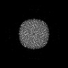 EMDB-45621: Apoferritin structure determined using laser phase contrast TEM (laser-off) |
| Source |
|
 Halobacillus (bacteria)