 Movie
Movie Controller
 Structure viewers
 About Yorodumi Papers


+Search query
-Structure paper
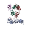
| Title | Effect of phosphorylation barcodes on arrestin binding to a chemokine receptor. |
|---|---|
| Journal, issue, pages | Nature, Vol. 643, Issue 8070, Page 280-287, Year 2025 |
| Publish date | May 21, 2025 |
 Authors Authors | Qiuyan Chen / Christopher T Schafer / Somnath Mukherjee / Kai Wang / Martin Gustavsson / James R Fuller / Katelyn Tepper / Thomas D Lamme / Yasmin Aydin / Parth Agrawal / Genki Terashi / Xin-Qiu Yao / Daisuke Kihara / Anthony A Kossiakoff / Tracy M Handel / John J G Tesmer /    |
| PubMed Abstract | Unique phosphorylation 'barcodes' installed in different regions of an active seven-transmembrane receptor by different G-protein-coupled receptor (GPCR) kinases (GRKs) have been proposed to promote ...Unique phosphorylation 'barcodes' installed in different regions of an active seven-transmembrane receptor by different G-protein-coupled receptor (GPCR) kinases (GRKs) have been proposed to promote distinct cellular outcomes, but it is unclear whether or how arrestins differentially engage these barcodes. Here, to address this, we developed an antigen-binding fragment (Fab7) that recognizes both active arrestin2 (β-arrestin1) and arrestin3 (β-arrestin2) without interacting with bound receptor polypeptides. We used Fab7 to determine the structures of both arrestins in complex with atypical chemokine receptor 3 (ACKR3) phosphorylated in different regions of its C-terminal tail by either GRK2 or GRK5 (ref. ). The GRK2-phosphorylated ACKR3 resulted in more heterogeneous 'tail-mode' assemblies, whereas phosphorylation by GRK5 resulted in more rigid 'ACKR3-adjacent' assemblies. Unexpectedly, the finger loops of both arrestins engaged the micelle surface rather than the receptor intracellular pocket, with arrestin3 being more dynamic, partly because of its lack of a membrane-anchoring motif. Thus, both the region of the barcode and the arrestin isoform involved can alter the structure and dynamics of GPCR-arrestin complexes, providing a possible mechanistic basis for unique downstream cellular effects, such as the efficiency of chemokine scavenging and the robustness of arrestin binding in ACKR3. |
 External links External links | Nature / PubMed:40399676 / PubMed Central |
| Methods | EM (single particle) |
| Resolution | 3.0 - 6.9 Å |
| Structure data | EMDB-41289, PDB-8tii:  EMDB-41290: Human ACKR3 phosphorylated by GRK5 in complex with Arrestin3  EMDB-41291: Human ACKR3 phosphorylated by GRK2 in complex with Arrestin3  EMDB-41292: Human ACKR3 with C tail extended by 12 glycines phosphorylated by GRK5 in complex with Arrestin2 EMDB-41295, PDB-8til: EMDB-41296, PDB-8tin: EMDB-41297, PDB-8tio: EMDB-43277: cryoEM structure of human ACKR3 phosphorylated by GRK5 in complex with Arrestin3 variant with the C edge loop from Arrestin2 inserted EMDB-47700, PDB-9e82:  EMDB-49564: cryoEM structure of human ACKR3 phosphorylated by GRK2 in complex with Arr2 |
| Chemicals |  ChemComp-CLR: |
| Source |
|
 Keywords Keywords | SIGNALING PROTEIN/IMMUNE SYSTEM / complex / GPCR / arrestin / signaling / SIGNALING PROTEIN-IMMUNE SYSTEM complex |


 homo sapiens (human)