Movie
Movie Controller
 Structure viewers
 About Yorodumi Papers


+Search query
-Structure paper
| Title | Tubulin acetyltransferases access and modify the microtubule luminal K40 residue through anchors in taxane-binding pockets. |
|---|---|
| Journal, issue, pages | Nat Struct Mol Biol, Vol. 32, Issue 2, Page 358-368, Year 2025 |
| Publish date | Nov 4, 2024 |
 Authors Authors | Jingyi Luo / Wai Hei Lam / Daqi Yu / Victor C Chao / Marc Nicholas Zopfi / Chen Jing Khoo / Chang Zhao / Shan Yan / Zheng Liu / Xiang David Li / Chaogu Zheng / Yuanliang Zhai / Shih-Chieh Ti /  |
| PubMed Abstract | Acetylation at α-tubulin K40 is the sole post-translational modification preferred to occur inside the lumen of hollow cylindrical microtubules. However, how tubulin acetyltransferases access the ...Acetylation at α-tubulin K40 is the sole post-translational modification preferred to occur inside the lumen of hollow cylindrical microtubules. However, how tubulin acetyltransferases access the luminal K40 in micrometer-long microtubules remains unknown. Here, we use cryo-electron microscopy and single-molecule reconstitution assays to reveal the enzymatic mechanism for tubulin acetyltransferases to modify K40 in the lumen. One tubulin acetyltransferase spans across the luminal lattice, with the catalytic core docking onto two α-tubulins and the enzyme's C-terminal domain occupying the taxane-binding pockets of two β-tubulins. The luminal accessibility and enzyme processivity of tubulin acetyltransferases are inhibited by paclitaxel, a microtubule-stabilizing chemotherapeutic agent. Characterizations using recombinant tubulins mimicking preacetylated and postacetylated K40 show the crosstalk between microtubule acetylation states and the cofactor acetyl-CoA in enzyme turnover. Our findings provide crucial insights into the conserved multivalent interactions involving α- and β-tubulins to acetylate the confined microtubule lumen. |
 External links External links | Nat Struct Mol Biol / PubMed:39496813 |
| Methods | EM (single particle) |
| Resolution | 3.1 - 3.6 Å |
| Structure data | EMDB-39076, PDB-8y9f: EMDB-39100, PDB-8yaj: EMDB-39102, PDB-8yal: EMDB-39105, PDB-8yar: |
| Chemicals |  ChemComp-GTP:  ChemComp-G2P: 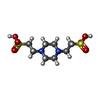 ChemComp-PIN:  ChemComp-ACO: |
| Source |
|
 Keywords Keywords | STRUCTURAL PROTEIN / Microtubules / luminal enzymes / PTMs / Microtubule / luminal enzyme / PTM |