[English] 日本語
Yorodumi Papers
- Database of articles cited by EMDB/PDB/SASBDB data -

+
Search query

Keywords
Structure methods
Author
Journal
IF

-
Structure paper

TitleMolecular mechanism of distinct chemokine engagement and functional divergence of the human Duffy antigen receptor.
Journal, issue, pagesCell, Vol. 187, Issue 17, Page 4751-4769.e25, Year 2024
Publish dateAug 22, 2024
AuthorsShirsha Saha / Basavraj Khanppnavar / Jagannath Maharana / Heeryung Kim / Carlo Marion C Carino / Carole Daly / Shane Houston / Saloni Sharma / Nashrah Zaidi / Annu Dalal / Sudha Mishra / Manisankar Ganguly / Divyanshu Tiwari / Poonam Kumari / Gagan Deep Jhingan / Prem N Yadav / Bianca Plouffe / Asuka Inoue / Ka Young Chung / Ramanuj Banerjee / Volodymyr M Korkhov / Arun K Shukla /
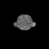
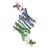
PubMed AbstractThe Duffy antigen receptor is a seven-transmembrane (7TM) protein expressed primarily at the surface of red blood cells and displays strikingly promiscuous binding to multiple inflammatory and ...The Duffy antigen receptor is a seven-transmembrane (7TM) protein expressed primarily at the surface of red blood cells and displays strikingly promiscuous binding to multiple inflammatory and homeostatic chemokines. It serves as the basis of the Duffy blood group system in humans and also acts as the primary attachment site for malarial parasite Plasmodium vivax and pore-forming toxins secreted by Staphylococcus aureus. Here, we comprehensively profile transducer coupling of this receptor, discover potential non-canonical signaling pathways, and determine the cryoelectron microscopy (cryo-EM) structure in complex with the chemokine CCL7. The structure reveals a distinct binding mode of chemokines, as reflected by relatively superficial binding and a partially formed orthosteric binding pocket. We also observe a dramatic shortening of TM5 and 6 on the intracellular side, which precludes the formation of the docking site for canonical signal transducers, thereby providing a possible explanation for the distinct pharmacological and functional phenotype of this receptor.
External linksCell / PubMed:39089252 / PubMed Central
MethodsEM (single particle)
Resolution3.65 - 4.23 Å
Structure data

EMDB-36488, PDB-8jps:
Structure of Duffy Antigen Receptor for Chemokines (DARC)/ACKR1 in complex with the chemokine, CCL7 (Composite map)
Method: EM (single particle) / Resolution: 3.65 Å

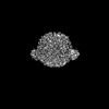
EMDB-37212: Structure of Duffy Antigen Receptor for Chemokines (DARC)/ACKR1 in complex with the chemokine, CCL7 (Receptor original map)
Method: EM (single particle) / Resolution: 3.65 Å

EMDB-37214: Structure of Duffy Antigen Receptor for Chemokines (DARC)/ACKR1 in complex with the chemokine, CCL7 (Ligand/CCL7 focused map)
Method: EM (single particle) / Resolution: 4.23 Å

Source
  • homo sapiens (human)
KeywordsIMMUNE SYSTEM / GPCR / SIGNALING PROTEIN

+
About Yorodumi Papers

-
News

-
Feb 9, 2022. New format data for meta-information of EMDB entries

New format data for meta-information of EMDB entries

  • Version 3 of the EMDB header file is now the official format.
  • The previous official version 1.9 will be removed from the archive.

Related info.:EMDB header

External links:wwPDB to switch to version 3 of the EMDB data model

-
Aug 12, 2020. Covid-19 info

Covid-19 info

URL: https://pdbjlvh1.pdbj.org/emnavi/covid19.php

New page: Covid-19 featured information page in EM Navigator.

Related info.:Covid-19 info / Mar 5, 2020. Novel coronavirus structure data

+
Mar 5, 2020. Novel coronavirus structure data

Novel coronavirus structure data

Related info.:Yorodumi Speices / Aug 12, 2020. Covid-19 info

External links:COVID-19 featured content - PDBj / Molecule of the Month (242):Coronavirus Proteases

+
Jan 31, 2019. EMDB accession codes are about to change! (news from PDBe EMDB page)

EMDB accession codes are about to change! (news from PDBe EMDB page)

  • The allocation of 4 digits for EMDB accession codes will soon come to an end. Whilst these codes will remain in use, new EMDB accession codes will include an additional digit and will expand incrementally as the available range of codes is exhausted. The current 4-digit format prefixed with “EMD-” (i.e. EMD-XXXX) will advance to a 5-digit format (i.e. EMD-XXXXX), and so on. It is currently estimated that the 4-digit codes will be depleted around Spring 2019, at which point the 5-digit format will come into force.
  • The EM Navigator/Yorodumi systems omit the EMD- prefix.

Related info.:Q: What is EMD? / ID/Accession-code notation in Yorodumi/EM Navigator

External links:EMDB Accession Codes are Changing Soon! / Contact to PDBj

+
Jul 12, 2017. Major update of PDB

Major update of PDB

  • wwPDB released updated PDB data conforming to the new PDBx/mmCIF dictionary.
  • This is a major update changing the version number from 4 to 5, and with Remediation, in which all the entries are updated.
  • In this update, many items about electron microscopy experimental information are reorganized (e.g. em_software).
  • Now, EM Navigator and Yorodumi are based on the updated data.

External links:wwPDB Remediation / Enriched Model Files Conforming to OneDep Data Standards Now Available in the PDB FTP Archive

-
Yorodumi Papers

Database of articles cited by EMDB/PDB/SASBDB data

  • Database of articles cited by EMDB, PDB, and SASBDB entries
  • Using PubMed data

Related info.:EMDB / PDB / SASBDB / Yorodumi / EMN Papers / Changes in new EM Navigator and Yorodumi

Read more