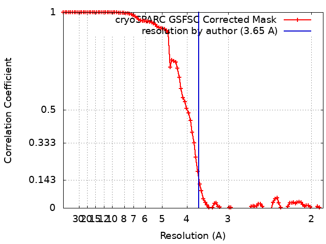
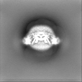
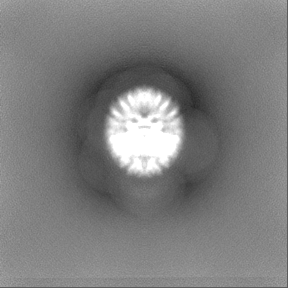
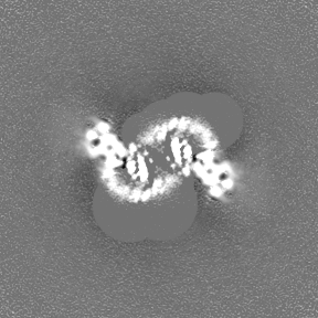
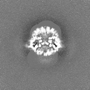
Journal: Cell / Year: 2024 Title: Molecular mechanism of distinct chemokine engagement and functional divergence of the human Duffy antigen receptor. Authors: Shirsha Saha / Basavraj Khanppnavar / Jagannath Maharana / Heeryung Kim / Carlo Marion C Carino / Carole Daly / Shane Houston / Saloni Sharma / Nashrah Zaidi / Annu Dalal / Sudha Mishra / ...Authors: Shirsha Saha / Basavraj Khanppnavar / Jagannath Maharana / Heeryung Kim / Carlo Marion C Carino / Carole Daly / Shane Houston / Saloni Sharma / Nashrah Zaidi / Annu Dalal / Sudha Mishra / Manisankar Ganguly / Divyanshu Tiwari / Poonam Kumari / Gagan Deep Jhingan / Prem N Yadav / Bianca Plouffe / Asuka Inoue / Ka Young Chung / Ramanuj Banerjee / Volodymyr M Korkhov / Arun K Shukla / Abstract: The Duffy antigen receptor is a seven-transmembrane (7TM) protein expressed primarily at the surface of red blood cells and displays strikingly promiscuous binding to multiple inflammatory and ...The Duffy antigen receptor is a seven-transmembrane (7TM) protein expressed primarily at the surface of red blood cells and displays strikingly promiscuous binding to multiple inflammatory and homeostatic chemokines. It serves as the basis of the Duffy blood group system in humans and also acts as the primary attachment site for malarial parasite Plasmodium vivax and pore-forming toxins secreted by Staphylococcus aureus. Here, we comprehensively profile transducer coupling of this receptor, discover potential non-canonical signaling pathways, and determine the cryoelectron microscopy (cryo-EM) structure in complex with the chemokine CCL7. The structure reveals a distinct binding mode of chemokines, as reflected by relatively superficial binding and a partially formed orthosteric binding pocket. We also observe a dramatic shortening of TM5 and 6 on the intracellular side, which precludes the formation of the docking site for canonical signal transducers, thereby providing a possible explanation for the distinct pharmacological and functional phenotype of this receptor.
Entire : Duffy Antigen Receptor for Chemokines (DARC)/ACKR1 in complex wit...
Entire
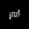
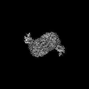
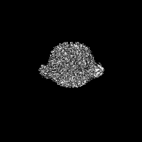
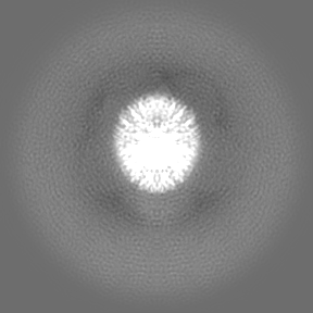
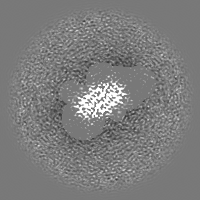
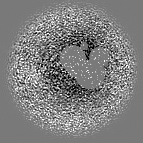
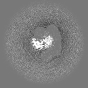
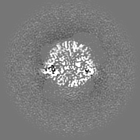
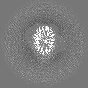
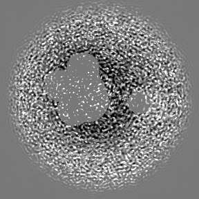
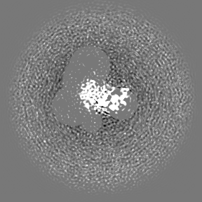
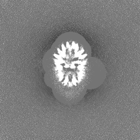
Name: Duffy Antigen Receptor for Chemokines (DARC)/ACKR1 in complex with the chemokine, CCL7
Components
Complex: Duffy Antigen Receptor for Chemokines (DARC)/ACKR1 in complex with the chemokine, CCL7
Complex: Duffy Antigen Receptor for Chemokines (DARC)/ACKR1
Protein or peptide: Atypical chemokine receptor 1
Complex: CCL7
Protein or peptide: C-C motif chemokine 7
-
Supramolecule #1: Duffy Antigen Receptor for Chemokines (DARC)/ACKR1 in complex wit...
Supramolecule
Name: Duffy Antigen Receptor for Chemokines (DARC)/ACKR1 in complex with the chemokine, CCL7 type: complex / ID: 1 / Parent: 0 / Macromolecule list: all
Source (natural)
Organism: Homo sapiens (human)
-
Supramolecule #2: Duffy Antigen Receptor for Chemokines (DARC)/ACKR1
In the structure databanks used in Yorodumi, some data are registered as the other names, "COVID-19 virus" and "2019-nCoV". Here are the details of the virus and the list of structure data.
Jan 31, 2019. EMDB accession codes are about to change! (news from PDBe EMDB page)
EMDB accession codes are about to change! (news from PDBe EMDB page)
The allocation of 4 digits for EMDB accession codes will soon come to an end. Whilst these codes will remain in use, new EMDB accession codes will include an additional digit and will expand incrementally as the available range of codes is exhausted. The current 4-digit format prefixed with “EMD-” (i.e. EMD-XXXX) will advance to a 5-digit format (i.e. EMD-XXXXX), and so on. It is currently estimated that the 4-digit codes will be depleted around Spring 2019, at which point the 5-digit format will come into force.
The EM Navigator/Yorodumi systems omit the EMD- prefix.
Related info.:Q: What is EMD? / ID/Accession-code notation in Yorodumi/EM Navigator
Yorodumi is a browser for structure data from EMDB, PDB, SASBDB, etc.
This page is also the successor to EM Navigator detail page, and also detail information page/front-end page for Omokage search.
The word "yorodu" (or yorozu) is an old Japanese word meaning "ten thousand". "mi" (miru) is to see.
Related info.:EMDB / PDB / SASBDB / Comparison of 3 databanks / Yorodumi Search / Aug 31, 2016. New EM Navigator & Yorodumi / Yorodumi Papers / Jmol/JSmol / Function and homology information / Changes in new EM Navigator and Yorodumi
 Movie
Movie Controller

 Yorodumi
 Open data
 Basic information
 Map data
 Sample
 Keywords
 Function and homology information
 Homo sapiens (human)
 Authors
 India,
  United Kingdom, 4 items
  Citation


 Structure visualization
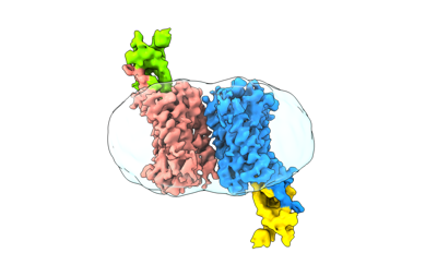
 Downloads & links
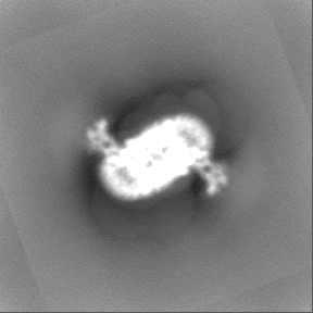
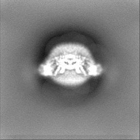
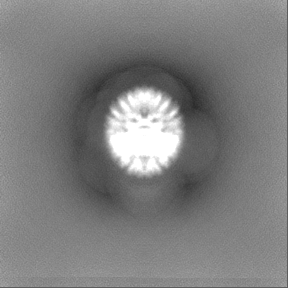
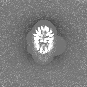
 emd_36488.png
 http://ftp.pdbj.org/pub/emdb/structures/EMD-36488


 Z (Sec.)
 Y (Row.)
 X (Col.)


 Sample components
 Spodoptera frugiperda (fall armyworm)
 Processing
 Electron microscopy
 FIELD EMISSION GUN