 Movie
Movie Controller
 Structure viewers
 About Yorodumi Papers


+Search query
-Structure paper
| Title | Nanoscale analysis of human G1 and metaphase chromatin in situ. |
|---|---|
| Journal, issue, pages | EMBO J, Vol. 44, Issue 9, Page 2658-2694, Year 2025 |
| Publish date | Mar 17, 2025 |
 Authors Authors | Jon Ken Chen / Tingsheng Liu / Shujun Cai / Weimei Ruan / Cai Tong Ng / Jian Shi / Uttam Surana / Lu Gan /   |
| PubMed Abstract | The structure of chromatin at the nucleosome level inside cells is still incompletely understood. Here we present in situ electron cryotomography analyses of chromatin in both G1 and metaphase RPE-1 ...The structure of chromatin at the nucleosome level inside cells is still incompletely understood. Here we present in situ electron cryotomography analyses of chromatin in both G1 and metaphase RPE-1 cells. G1 nucleosomes are concentrated in globular chromatin domains, and metaphase nucleosomes are concentrated in the chromatids. Classification analysis reveals that canonical mononucleosomes, and in some conditions ordered stacked dinucleosomes and mononucleosomes with a disordered gyre-proximal density, are abundant in both cell-cycle states. We do not detect class averages that have more than two stacked nucleosomes or side-by-side dinucleosomes, suggesting that groups of more than two nucleosomes are heterogeneous. Large multi-megadalton structures are abundant in G1 nucleoplasm, but not found in G1 chromatin domains and metaphase chromatin. The macromolecular phenotypes studied here represent a starting point for the comparative analysis of compaction in normal vs. unhealthy human cells, in other cell-cycle states, other organisms, and in vitro chromatin assemblies. |
 External links External links | EMBO J / PubMed:40097852 / PubMed Central |
| Methods | EM (subtomogram averaging) |
| Resolution | 24.0 - 36.8 Å |
| Structure data |  EMDB-36992: G1 dinucleosome  EMDB-36993: G1 mononucleosome  EMDB-36994: G1 nucleosome gyre proximal  EMDB-36998: Metaphase dinucleosome  EMDB-36999: Metaphase mononucleosome  EMDB-37000: Metaphase nucleosome proximal  EMDB-37001: G1 preribosome  EMDB-37002: G1 ribosome cytoplasmic  EMDB-37003: Metaphase ribosome 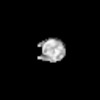 EMDB-37004: Oligonucleosome 0 DMSO  EMDB-37005: Oligonucleosome 9% DMSO 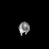 EMDB-62350: G1 mononucleosome, no cryoprotectant 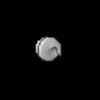 EMDB-62351: G1 mononucleosome, with glycerol cryoprotectant 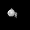 EMDB-62352: G1 nucleosome with gyre proximal density, with glycerol cryoprotectant |
| Source |
|
 Homo sapiens (human)