 Movie
Movie Controller
 Structure viewers
 About Yorodumi Papers


+Search query
-Structure paper
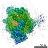
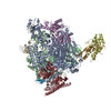
| Title | Structure of transcribing mammalian RNA polymerase II. |
|---|---|
| Journal, issue, pages | Nature, Vol. 529, Issue 7587, Page 551-554, Year 2016 |
| Publish date | Jan 28, 2016 |
 Authors Authors | Carrie Bernecky / Franz Herzog / Wolfgang Baumeister / Jürgen M Plitzko / Patrick Cramer /  |
| PubMed Abstract | RNA polymerase (Pol) II produces messenger RNA during transcription of protein-coding genes in all eukaryotic cells. The Pol II structure is known at high resolution from X-ray crystallography for ...RNA polymerase (Pol) II produces messenger RNA during transcription of protein-coding genes in all eukaryotic cells. The Pol II structure is known at high resolution from X-ray crystallography for two yeast species. Structural studies of mammalian Pol II, however, remain limited to low-resolution electron microscopy analysis of human Pol II and its complexes with various proteins. Here we report the 3.4 Å resolution cryo-electron microscopy structure of mammalian Pol II in the form of a transcribing complex comprising DNA template and RNA transcript. We use bovine Pol II, which is identical to the human enzyme except for seven amino-acid residues. The obtained atomic model closely resembles its yeast counterpart, but also reveals unknown features. Binding of nucleic acids to the polymerase involves 'induced fit' of the mobile Pol II clamp and active centre region. DNA downstream of the transcription bubble contacts a conserved 'TPSA motif' in the jaw domain of the Pol II subunit RPB5, an interaction that is apparently already established during transcription initiation. Upstream DNA emanates from the active centre cleft at an angle of approximately 105° with respect to downstream DNA. This position of upstream DNA allows for binding of the general transcription elongation factor DSIF (SPT4-SPT5) that we localize over the active centre cleft in a conserved position on the clamp domain of Pol II. Our results define the structure of mammalian Pol II in its functional state, indicate that previous crystallographic analysis of yeast Pol II is relevant for understanding gene transcription in all eukaryotes, and provide a starting point for a mechanistic analysis of human transcription. |
 External links External links | Nature / PubMed:26789250 |
| Methods | EM (single particle) |
| Resolution | 3.4 - 3.7 Å |
| Structure data | EMDB-3218: Structure of transcribing mammalian RNA polymerase II (EC1) 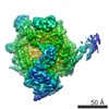 EMDB-3219:  EMDB-3220: |
| Chemicals |  ChemComp-ZN:  ChemComp-MG: |
| Source |
|
 Keywords Keywords | TRANSCRIPTION / ELONGATION |

 Homo sapiens (human)