 Movie
Movie Controller
 Structure viewers
 About Yorodumi Papers


+Search query
-Structure paper
| Title | Cryogenic single-molecule fluorescence annotations for electron tomography reveal in situ organization of key proteins in . |
|---|---|
| Journal, issue, pages | Proc Natl Acad Sci U S A, Vol. 117, Issue 25, Page 13937-13944, Year 2020 |
| Publish date | Jun 23, 2020 |
 Authors Authors | Peter D Dahlberg / Saumya Saurabh / Annina M Sartor / Jiarui Wang / Patrick G Mitchell / Wah Chiu / Lucy Shapiro / W E Moerner /  |
| PubMed Abstract | Superresolution fluorescence microscopy and cryogenic electron tomography (CET) are powerful imaging methods for exploring the subcellular organization of biomolecules. Superresolution fluorescence ...Superresolution fluorescence microscopy and cryogenic electron tomography (CET) are powerful imaging methods for exploring the subcellular organization of biomolecules. Superresolution fluorescence microscopy based on covalent labeling highlights specific proteins and has sufficient sensitivity to observe single fluorescent molecules, but the reconstructions lack detailed cellular context. CET has molecular-scale resolution but lacks specific and nonperturbative intracellular labeling techniques. Here, we describe an imaging scheme that correlates cryogenic single-molecule fluorescence localizations with CET reconstructions. Our approach achieves single-molecule localizations with an average lateral precision of 9 nm, and a relative registration error between the set of localizations and CET reconstruction of ∼30 nm. We illustrate the workflow by annotating the positions of three proteins in the bacterium : McpA, PopZ, and SpmX. McpA, which forms a part of the chemoreceptor array, acts as a validation structure by being visible under both imaging modalities. In contrast, PopZ and SpmX cannot be directly identified in CET. While not directly discernable, PopZ fills a region at the cell poles that is devoid of electron-dense ribosomes. We annotate the position of PopZ with single-molecule localizations and confirm its position within the ribosome excluded region. We further use the locations of PopZ to provide context for localizations of SpmX, a low-copy integral membrane protein sequestered by PopZ as part of a signaling pathway that leads to an asymmetric cell division. Our correlative approach reveals that SpmX localizes along one side of the cell pole and its extent closely matches that of the PopZ region. |
 External links External links | Proc Natl Acad Sci U S A / PubMed:32513734 / PubMed Central |
| Methods | EM (tomography) |
| Structure data | 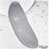 EMDB-21706: |
| Source |
|
 Caulobacter vibrioides (bacteria)