Movie
Movie Controller
 Structure viewers
 About Yorodumi Papers


+Search query
-Structure paper
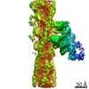
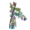
| Title | The molecular basis for sarcomere organization in vertebrate skeletal muscle. |
|---|---|
| Journal, issue, pages | Cell, Vol. 184, Issue 8, Page 2135-2150.e13, Year 2021 |
| Publish date | Apr 15, 2021 |
 Authors Authors | Zhexin Wang / Michael Grange / Thorsten Wagner / Ay Lin Kho / Mathias Gautel / Stefan Raunser /   |
| PubMed Abstract | Sarcomeres are force-generating and load-bearing devices of muscles. A precise molecular picture of how sarcomeres are built underpins understanding their role in health and disease. Here, we ...Sarcomeres are force-generating and load-bearing devices of muscles. A precise molecular picture of how sarcomeres are built underpins understanding their role in health and disease. Here, we determine the molecular architecture of native vertebrate skeletal sarcomeres by electron cryo-tomography. Our reconstruction reveals molecular details of the three-dimensional organization and interaction of actin and myosin in the A-band, I-band, and Z-disc and demonstrates that α-actinin cross-links antiparallel actin filaments by forming doublets with 6-nm spacing. Structures of myosin, tropomyosin, and actin at ~10 Å further reveal two conformations of the "double-head" myosin, where the flexible orientation of the lever arm and light chains enable myosin not only to interact with the same actin filament, but also to split between two actin filaments. Our results provide unexpected insights into the fundamental organization of vertebrate skeletal muscle and serve as a strong foundation for future investigations of muscle diseases. |
 External links External links | Cell / PubMed:33765442 / PubMed Central |
| Methods | EM (subtomogram averaging) |
| Resolution | 10.2 - 19.8 Å |
| Structure data | EMDB-12289: Structure of the in situ actomyosin complex from the A-band of mouse psoas muscle sarcomere in the rigor state obtained by sub-tomogram averaging 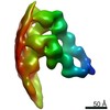 EMDB-12291: 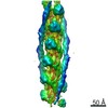 EMDB-12292: 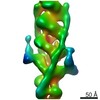 EMDB-12293: |
| Source |
|
 Keywords Keywords | MOTOR PROTEIN / Muscle proteins / force generation / sarcomere / cytoskeleton |