 Movie
Movie Controller
 Structure viewers
 About Yorodumi Papers


+Search query
-Structure paper
| Title | Elongated oligomers assemble into mammalian PrP amyloid fibrils. |
|---|---|
| Journal, issue, pages | J Mol Biol, Vol. 357, Issue 3, Page 975-985, Year 2006 |
| Publish date | Mar 31, 2006 |
 Authors Authors | M Howard Tattum / Sara Cohen-Krausz / Kanjana Thumanu / Christopher W Wharton / Azadeh Khalili-Shirazi / Graham S Jackson / Elena V Orlova / John Collinge / Anthony R Clarke / Helen R Saibil /  |
| PubMed Abstract | In prion diseases, the mammalian prion protein PrP is converted from a monomeric, mainly alpha-helical state into beta-rich amyloid fibrils. To examine the structure of the misfolded state, amyloid ...In prion diseases, the mammalian prion protein PrP is converted from a monomeric, mainly alpha-helical state into beta-rich amyloid fibrils. To examine the structure of the misfolded state, amyloid fibrils were grown from a beta form of recombinant mouse PrP (residues 91-231). The beta-PrP precursors assembled slowly into amyloid fibrils with an overall helical twist. The fibrils exhibit immunological reactivity similar to that of ex vivo PrP Sc. Using electron microscopy and image processing, we obtained three-dimensional density maps of two forms of PrP fibrils with slightly different twists. They reveal two intertwined protofilaments with a subunit repeat of approximately 60 A. The repeating unit along each protofilament can be accounted for by elongated oligomers of PrP, suggesting a hierarchical assembly mechanism for the fibrils. The structure reveals flexible crossbridges between the two protofilaments, and subunit contacts along the protofilaments that are likely to reflect specific features of the PrP sequence, in addition to the generic, cross-beta amyloid fold. |
 External links External links | J Mol Biol / PubMed:16473369 |
| Methods | EM (single particle) |
| Resolution | 26.0 - 28.0 Å |
| Structure data | 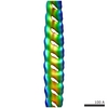 EMDB-1186:  EMDB-1187: |
| Source |
|

