 Movie
Movie Controller
 Structure viewers
 About Yorodumi Papers


+Search query
-Structure paper
| Title | CryoEM at 100 keV: a demonstration and prospects. |
|---|---|
| Journal, issue, pages | IUCrJ, Vol. 6, Issue Pt 6, Page 1086-1098, Year 2019 |
| Publish date | Nov 1, 2019 |
 Authors Authors | K Naydenova / G McMullan / M J Peet / Y Lee / P C Edwards / S Chen / E Leahy / S Scotcher / R Henderson / C J Russo /  |
| PubMed Abstract | 100 kV is investigated as the operating voltage for single-particle electron cryomicroscopy (cryoEM). Reducing the electron energy from the current standard of 300 or 200 keV offers both cost ...100 kV is investigated as the operating voltage for single-particle electron cryomicroscopy (cryoEM). Reducing the electron energy from the current standard of 300 or 200 keV offers both cost savings and potentially improved imaging. The latter follows from recent measurements of radiation damage to biological specimens by high-energy electrons, which show that at lower energies there is an increased amount of information available per unit damage. For frozen hydrated specimens around 300 Å in thickness, the predicted optimal electron energy for imaging is 100 keV. Currently available electron cryomicroscopes in the 100-120 keV range are not optimized for cryoEM as they lack both the spatially coherent illumination needed for the high defocus used in cryoEM and imaging detectors optimized for 100 keV electrons. To demonstrate the potential of imaging at 100 kV, the voltage of a standard, commercial 200 kV field-emission gun (FEG) microscope was reduced to 100 kV and a side-entry cryoholder was used. As high-efficiency, large-area cameras are not currently available for 100 keV electrons, a commercial hybrid pixel camera designed for X-ray detection was attached to the camera chamber and was used for low-dose data collection. Using this configuration, five single-particle specimens were imaged: hepatitis B virus capsid, bacterial 70S ribosome, catalase, DNA protection during starvation protein and haemoglobin, ranging in size from 4.5 MDa to 64 kDa with corresponding diameters from 320 to 72 Å. These five data sets were used to reconstruct 3D structures with resolutions between 8.4 and 3.4 Å. Based on this work, the practical advantages and current technological limitations to single-particle cryoEM at 100 keV are considered. These results are also discussed in the context of future microscope development towards the goal of rapid, simple and widely available structure determination of any purified biological specimen. |
 External links External links | IUCrJ / PubMed:31709064 / PubMed Central |
| Methods | EM (single particle) |
| Resolution | 3.4 - 7.0 Å |
| Structure data | 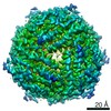 EMDB-10161:  EMDB-10265: |
| Source |
|

