[English] 日本語
Yorodumi Papers
- Database of articles cited by EMDB/PDB/SASBDB data -

+
Search query

Keywords
Structure methods
Author
Journal
IF

-
Structure paper

TitleUsing stable MutS dimers and tetramers to quantitatively analyze DNA mismatch recognition and sliding clamp formation.
Journal, issue, pagesNucleic Acids Res, Vol. 41, Issue 17, Page 8166-8181, Year 2013
Publish dateJul 1, 2013
AuthorsFlora S Groothuizen / Alexander Fish / Maxim V Petoukhov / Annet Reumer / Laura Manelyte / Herrie H K Winterwerp / Martin G Marinus / Joyce H G Lebbink / Dmitri I Svergun / Peter Friedhoff / Titia K Sixma /
PubMed AbstractThe process of DNA mismatch repair is initiated when MutS recognizes mismatched DNA bases and starts the repair cascade. The Escherichia coli MutS protein exists in an equilibrium between dimers and ...The process of DNA mismatch repair is initiated when MutS recognizes mismatched DNA bases and starts the repair cascade. The Escherichia coli MutS protein exists in an equilibrium between dimers and tetramers, which has compromised biophysical analysis. To uncouple these states, we have generated stable dimers and tetramers, respectively. These proteins allowed kinetic analysis of DNA recognition and structural analysis of the full-length protein by X-ray crystallography and small angle X-ray scattering. Our structural data reveal that the tetramerization domains are flexible with respect to the body of the protein, resulting in mostly extended structures. Tetrameric MutS has a slow dissociation from DNA, which can be due to occasional bending over and binding DNA in its two binding sites. In contrast, the dimer dissociation is faster, primarily dependent on a combination of the type of mismatch and the flanking sequence. In the presence of ATP, we could distinguish two kinetic groups: DNA sequences where MutS forms sliding clamps and those where sliding clamps are not formed efficiently. Interestingly, this inability to undergo a conformational change rather than mismatch affinity is correlated with mismatch repair.
External linksNucleic Acids Res / PubMed:23821665 / PubMed Central
MethodsSAS (X-ray synchrotron) / X-ray diffraction
Resolution3.1 Å
Structure data

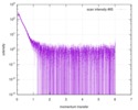
SASDA24: MutS tetramer (DNA mismatch repair protein MutS)
Method: SAXS/SANS

SASDAN3:
MutS dimer (DNA mismatch repair protein MutS, MutS)
Method: SAXS/SANS

SASDAQ3:
MutS tetramer (DNA mismatch repair protein MutS)
Method: SAXS/SANS

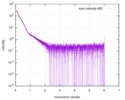
SASDAX3: MutS tetramer (DNA mismatch repair protein MutS)
Method: SAXS/SANS

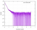
SASDAY3: MutS tetramer (DNA mismatch repair protein MutS)
Method: SAXS/SANS

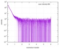
SASDAZ3: MutS tetramer (DNA mismatch repair protein MutS)
Method: SAXS/SANS

PDB-3zlj:
CRYSTAL STRUCTURE OF FULL-LENGTH E.COLI DNA MISMATCH REPAIR PROTEIN MUTS D835R MUTANT IN COMPLEX WITH GT MISMATCHED DNA
Method: X-RAY DIFFRACTION / Resolution: 3.1 Å

Source
  • Escherichia coli (E. coli)
  • escherichia coli k-12 (bacteria)
  • synthetic construct (others)
KeywordsDNA BINDING PROTEIN/DNA / DNA BINDING PROTEIN-DNA COMPLEX / DIMER MUTANT / MISMATCH REPAIR / DNA REPAIR PROTEIN / DNA DAMAGE / NUCLEOTIDE-BINDING / ATP-BINDING

+
About Yorodumi Papers

-
News

-
Feb 9, 2022. New format data for meta-information of EMDB entries

New format data for meta-information of EMDB entries

  • Version 3 of the EMDB header file is now the official format.
  • The previous official version 1.9 will be removed from the archive.

Related info.:EMDB header

External links:wwPDB to switch to version 3 of the EMDB data model

-
Aug 12, 2020. Covid-19 info

Covid-19 info

URL: https://pdbjlvh1.pdbj.org/emnavi/covid19.php

New page: Covid-19 featured information page in EM Navigator.

Related info.:Covid-19 info / Mar 5, 2020. Novel coronavirus structure data

+
Mar 5, 2020. Novel coronavirus structure data

Novel coronavirus structure data

Related info.:Yorodumi Speices / Aug 12, 2020. Covid-19 info

External links:COVID-19 featured content - PDBj / Molecule of the Month (242):Coronavirus Proteases

+
Jan 31, 2019. EMDB accession codes are about to change! (news from PDBe EMDB page)

EMDB accession codes are about to change! (news from PDBe EMDB page)

  • The allocation of 4 digits for EMDB accession codes will soon come to an end. Whilst these codes will remain in use, new EMDB accession codes will include an additional digit and will expand incrementally as the available range of codes is exhausted. The current 4-digit format prefixed with “EMD-” (i.e. EMD-XXXX) will advance to a 5-digit format (i.e. EMD-XXXXX), and so on. It is currently estimated that the 4-digit codes will be depleted around Spring 2019, at which point the 5-digit format will come into force.
  • The EM Navigator/Yorodumi systems omit the EMD- prefix.

Related info.:Q: What is EMD? / ID/Accession-code notation in Yorodumi/EM Navigator

External links:EMDB Accession Codes are Changing Soon! / Contact to PDBj

+
Jul 12, 2017. Major update of PDB

Major update of PDB

  • wwPDB released updated PDB data conforming to the new PDBx/mmCIF dictionary.
  • This is a major update changing the version number from 4 to 5, and with Remediation, in which all the entries are updated.
  • In this update, many items about electron microscopy experimental information are reorganized (e.g. em_software).
  • Now, EM Navigator and Yorodumi are based on the updated data.

External links:wwPDB Remediation / Enriched Model Files Conforming to OneDep Data Standards Now Available in the PDB FTP Archive

-
Yorodumi Papers

Database of articles cited by EMDB/PDB/SASBDB data

  • Database of articles cited by EMDB, PDB, and SASBDB entries
  • Using PubMed data

Related info.:EMDB / PDB / SASBDB / Yorodumi / EMN Papers / Changes in new EM Navigator and Yorodumi

Read more