 ムービー
ムービー コントローラー
 構造ビューア
 万見文献について


+検索条件
-Structure paper
| タイトル | Non-equilibrium snapshots of ligand efficacy at the μ-opioid receptor. |
|---|---|
| ジャーナル・号・ページ | Nature, Year 2025 |
| 掲載日 | 2025年12月22日 |
 著者 著者 | Michael J Robertson / Arnab Modak / Makaía M Papasergi-Scott / Miaohui Hu / Maria Claudia Peroto / Balazs R Varga / Susruta Majumdar / Ravi Kalathur / Scott C Blanchard / Georgios Skiniotis /  |
| PubMed 要旨 | Distinct ligands for the same G-protein-coupled receptor (GPCR) activate intracellular signalling partners to varying extents, but the molecular mechanisms that drive these differences remain elusive. ...Distinct ligands for the same G-protein-coupled receptor (GPCR) activate intracellular signalling partners to varying extents, but the molecular mechanisms that drive these differences remain elusive. Here, hypothesizing that such differences in signalling efficacy might be captured structurally in intermediate states under non-equilibrium conditions, we use a time-resolved cryo-electron-microscopy approach to visualize the GTP-induced activation of the Gαβγ heterotrimer by the μ-opioid receptor bound to three ligands that show partial, full or super agonism on the receptor. We resolve ensembles of conformational states along the G-protein activation pathway, including an intermediate state that enables us to visualize receptor dynamics as a function of bound ligand. The results reveal ligand-dependent differences in state occupancy and conformational stability, with higher ligand efficacy correlating with increased dynamics of the receptor's transmembrane helices 5 and 6. Furthermore, we identify key differences between G and G in the mechanism of GTP-induced activation, which are likely to underlie the distinct activation kinetics of these G-protein types. Corroborated by molecular-dynamics simulations and single-molecule fluorescence assays, our findings provide a dynamic structural landscape of GPCR-G-protein interactions for ligands of various efficacies, and suggest that partial agonists produce a 'kinetic trap' during G-protein activation. |
 リンク リンク | Nature / PubMed:41430437 |
| 手法 | EM (単粒子) |
| 解像度 | 2.4 - 6.5 Å |
| 構造データ | EMDB-70356, PDB-9ode: EMDB-70357, PDB-9odf: EMDB-70358, PDB-9odg: EMDB-70359, PDB-9odh: EMDB-70360, PDB-9odi:  EMDB-70361: Structure of the MOR/Gi/Lofentanil Complex, GTP-bound G-ACT-2/3, Global and G Protein Local  EMDB-70362: Structure of the MOR/Gi/Lofentanil Complex, GTP-bound G-ACT-2/3, Global 3DVA Sorted 1  EMDB-70363: Structure of the MOR/Gi/Lofentanil Complex, GTP-bound G-ACT-2/3, Global 3DVA Sorted 2 EMDB-70364, PDB-9odj: EMDB-70365, PDB-9odk: EMDB-70366, PDB-9odl:  EMDB-70373: Structure of the MOR/Gi/DAMGO Complex, GTP-Bound, G-ACT-1  EMDB-70374: Structure of the MOR/Gi/DAMGO Complex, GTP-Bound, G-ACT-2/3 Consensus Refinement |
| 化合物 | 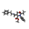 ChemComp-EID:  ChemComp-GTP:  ChemComp-MG:  ChemComp-CLR:  ChemComp-EIG: |
| 由来 |
|
 キーワード キーワード | MEMBRANE PROTEIN / GPCR / Complex / Agonist |


 homo sapiens (ヒト)
