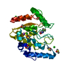
Crystal structures of non-uracil ring fragments in complex with Mycobacterium tuberculosis uracil DNA glycosylase (MtUng) as a starting point for novel inhibitor design: A case study with the barbituric acid fragment.
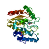
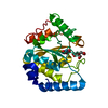
PDB-8i61: Crystal structure of Mycobacterium tuberculosis Uracil-DNA glycosylase in complex with Barbituric acid and Citric acid, Form I 手法: X-RAY DIFFRACTION / 解像度: 1.24 Å
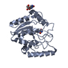
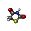
PDB-8i62: Crystal structure of Mycobacterium tuberculosis Uracil-DNA glycosylase in complex with Barbituric acid, Form I 手法: X-RAY DIFFRACTION / 解像度: 1.26 Å
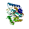
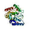
PDB-8i63: Crystal structure of Mycobacterium tuberculosis Uracil-DNA glycosylase in complex with Barbituric acid, Form III 手法: X-RAY DIFFRACTION / 解像度: 1.95 Å
PDB-8i64: Crystal structure of Mycobacterium tuberculosis Uracil-DNA glycosylase in complex with Barbituric acid, Form II 手法: X-RAY DIFFRACTION / 解像度: 2.26 Å
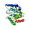
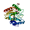
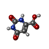
PDB-8i65: Crystal structure of Mycobacterium tuberculosis Uracil-DNA glycosylase in complex with isoorotic acid (2,4-Dihydroxypyrimidine-5-carboxylic Acid), Form I 手法: X-RAY DIFFRACTION / 解像度: 1.72 Å
PDB-8i66: Crystal structure of Mycobacterium tuberculosis Uracil-DNA glycosylase in complex with isoorotic acid (2,4-Dihydroxypyrimidine-5-carboxylic Acid) and citric acid, Form I 手法: X-RAY DIFFRACTION / 解像度: 2.6 Å
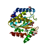
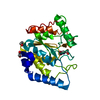
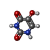
PDB-8i67: Crystal structure of Mycobacterium tuberculosis Uracil-DNA glycosylase in complex with 2,4-Thiazolidinedione, Form I 手法: X-RAY DIFFRACTION / 解像度: 1.72 Å
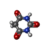
PDB-8i68: Crystal structure of Mycobacterium tuberculosis Uracil-DNA glycosylase in complex with Uric acid, Form III 手法: X-RAY DIFFRACTION / 解像度: 1.88 Å
PDB-8i69: Crystal structure of Mycobacterium tuberculosis Uracil-DNA glycosylase in complex with 5-Fluoroorotic acid and Citric acid, Form I 手法: X-RAY DIFFRACTION / 解像度: 2 Å
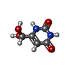
PDB-8i6a: Crystal structure of Mycobacterium tuberculosis Uracil-DNA glycosylase in complex with Orotic acid, Form III 手法: X-RAY DIFFRACTION / 解像度: 2 Å
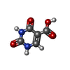
PDB-8i6b: Crystal structure of Mycobacterium tuberculosis Uracil-DNA glycosylase in complex with 5-Hydroxy-2,4(1H,3H)-pyrimidinedione, Form I 手法: X-RAY DIFFRACTION / 解像度: 1.6 Å
PDB-8i6c: Crystal structure of Mycobacterium tuberculosis Uracil-DNA glycosylase in complex with 6-Formyl-uracil, Form III 手法: X-RAY DIFFRACTION / 解像度: 2.28 Å
PDB-8i6d: Crystal structure of Mycobacterium tuberculosis Uracil-DNA glycosylase in complex with 5-Hydroxy-2,4(1H,3H)-pyrimidinedione, Form VI 手法: X-RAY DIFFRACTION / 解像度: 2.4 Å
 ムービー
ムービー コントローラー
 構造ビューア
 万見文献について


 著者
著者 リンク
リンク


 キーワード
キーワード mycobacterium tuberculosis h37rv (結核菌)