 Movie
Movie Controller


+ Open data

- Basic information
| Entry |  Database: PDB chemical components / ID: 5CU Database: PDB chemical components / ID: 5CU |
|---|---|
| Name | Name: |
-Chemical information
| Composition |  | ||||
|---|---|---|---|---|---|
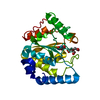
| Others | Type: NON-POLYMER / PDB classification: HETAIN / Three letter code: 5CU / Ideal coordinates details: Corina / Model coordinates PDB-ID: 4LAL | ||||
| History |
| ||||
 External links External links | UniChem / ChemSpider / Brenda / ChEBI / ChEMBL / CompTox / PubChem / SureChEMBL / Wikipedia search / Google search |
- Structure visualization
| Structure viewer | Molecule:  MolmilJmol/JSmol MolmilJmol/JSmol |
|---|
-Details
-SMILES
| ACDLabs 12.01 | | CACTVS 3.385 | OpenEye OEToolkits 1.7.6 | |
|---|
-SMILES CANONICAL
| CACTVS 3.385 | | OpenEye OEToolkits 1.7.6 | |
|---|
-InChI
| InChI 1.03 |
|---|
-InChIKey
| InChI 1.03 |
|---|
-SYSTEMATIC NAME
| ACDLabs 12.01 | | OpenEye OEToolkits 1.7.6 | |
|---|
-PDB entries
Showing all 4 items
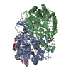
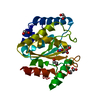
PDB-4lal: 
Crystal structure of Cordyceps militaris IDCase D323A mutant in complex with 5-carboxyl-uracil
PDB-4lam:
Crystal structure of Cordyceps militaris IDCase D323N mutant in complex with 5-carboxyl-uracil
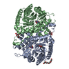
PDB-8i65:
Crystal structure of Mycobacterium tuberculosis Uracil-DNA glycosylase in complex with isoorotic acid (2,4-Dihydroxypyrimidine-5-carboxylic Acid), Form I
PDB-8i66:
Crystal structure of Mycobacterium tuberculosis Uracil-DNA glycosylase in complex with isoorotic acid (2,4-Dihydroxypyrimidine-5-carboxylic Acid) and citric acid, Form I
