 ムービー
ムービー コントローラー
 構造ビューア
 万見文献について


+検索条件
-Structure paper
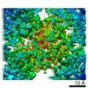
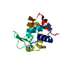
| タイトル | Using focus ion beam to prepare crystal lamella for electron diffraction. |
|---|---|
| ジャーナル・号・ページ | J Struct Biol, Vol. 205, Issue 3, Page 59-64, Year 2019 |
| 掲載日 | 2019年3月1日 |
 著者 著者 | Heng Zhou / Zhipu Luo / Xueming Li /  |
| PubMed 要旨 | Electron diffraction provides a powerful tool to solve the structures of small protein crystals. However, strong interactions between the electrons and the materials limit the application of the ...Electron diffraction provides a powerful tool to solve the structures of small protein crystals. However, strong interactions between the electrons and the materials limit the application of the electron crystallographic method on large protein crystals with micrometer or larger sizes. Here, we used the focused ion beam (FIB) equipped on the scanning electron microscope (SEM) to mill a large crystal to thin lamella. The influences of the milling on the crystal lamella were observed and investigated, including radiation damage on the crystal surface during the FIB imaging, deformation of the thin crystal lamella, and variation in the diffraction intensities under electron radiation. These observations provide important information to optimize the FIB milling, and hence is important to obtain high-quality crystal samples for routine structure determination of protein crystals using the electron cryo-microscope. |
 リンク リンク | J Struct Biol / PubMed:30794865 |
| 手法 | EM (電子線結晶学) |
| 解像度 | 1.5 - 1.73 Å |
| 構造データ | |
| 化合物 |  ChemComp-ACT:  ChemComp-HOH:  ChemComp-SO4: |
| 由来 |
|
 キーワード キーワード | HYDROLASE / Lysozyme / MicroED / Focused Ion Beam / crystal lamella / Proteinase K |


 parengyodontium album (菌類)