| タイトル | Structural basis of Dscam1 homodimerization: Insights into context constraint for protein recognition |
|---|
| ジャーナル・号・ページ | Sci Adv, Vol. 2, Page e1501118-e1501118, Year 2016 |
|---|
| 掲載日 | 2014年11月7日 (構造データの登録日) |
|---|
 著者 著者 | Li, S.A. / Cheng, L. / Yu, Y. / Chen, Q. |
|---|
 リンク リンク |  Sci Adv / Sci Adv /  PubMed:27386517 PubMed:27386517 |
|---|
| 手法 | X線回折 |
|---|
| 解像度 | 1.902 - 4.004 Å |
|---|
| 構造データ | PDB-4wvr:
Crystal structure of Dscam1 Ig7 domain, isoform 5
手法: X-RAY DIFFRACTION / 解像度: 1.948 Å PDB-4x5l:
Crystal structure of Dscam1 Ig7 domain, isoform 9
手法: X-RAY DIFFRACTION / 解像度: 2.374 Å PDB-4x83:
Crystal structure of Dscam1 isoform 7.44, N-terminal four Ig domains
手法: X-RAY DIFFRACTION / 解像度: 1.902 Å PDB-4x8x:
Crystal structure of Dscam1 isoform 1.9, N-terminal four Ig domains
手法: X-RAY DIFFRACTION / 解像度: 2.5 Å PDB-4x9b:
Crystal structure of Dscam1 isoform 4.44, N-terminal four Ig domains
手法: X-RAY DIFFRACTION / 解像度: 2.2 Å PDB-4x9f:
Crystal structure of Dscam1 isoform 6.9, N-terminal four Ig domains
手法: X-RAY DIFFRACTION / 解像度: 2.35 Å PDB-4x9g:
Crystal structure of Dscam1 isoform 6.44, N-terminal four Ig domains
手法: X-RAY DIFFRACTION / 解像度: 3.403 Å PDB-4x9h:
Crystal structure of Dscam1 isoform 8.4, N-terminal four Ig domains
手法: X-RAY DIFFRACTION / 解像度: 2.95 Å PDB-4x9i:
Crystal structure of Dscam1 isoform 9.44, N-terminal four Ig domains
手法: X-RAY DIFFRACTION / 解像度: 2.904 Å PDB-4xb7:
Crystal structure of Dscam1 isoform 4.4, N-terminal four Ig domains
手法: X-RAY DIFFRACTION / 解像度: 4.004 Å PDB-4xb8:
Crystal structure of Dscam1 isoform 9.44, N-terminal four Ig domains (with zinc)
手法: X-RAY DIFFRACTION / 解像度: 3.202 Å PDB-4xhq:
Re-refinement the crystal structure of Dscam1 isoform 1.34, N-terminal four Ig domains
手法: X-RAY DIFFRACTION / 解像度: 1.948 Å |
|---|
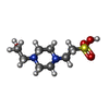
| 化合物 | ChemComp-EPE:
4-(2-HYDROXYETHYL)-1-PIPERAZINE ETHANESULFONIC ACID / HEPES / pH緩衝剤*YM
|
|---|
| 由来 |   drosophila melanogaster (キイロショウジョウバエ) drosophila melanogaster (キイロショウジョウバエ)
|
|---|
 キーワード キーワード | CELL ADHESION / Ig fold |
|---|
 著者
著者 リンク
リンク Sci Adv /
  PubMed:27386517


 キーワード
キーワード ムービー
ムービー コントローラー
 構造ビューア
 万見文献について


