PDB-4d8v: Crystal structure of the hexameric purine nucleoside phosphorylase from Bacillus subtilis at pH 4.2 手法: X-RAY DIFFRACTION / 解像度: 2.35 Å
PDB-4d8x: Crystal structure of the hexameric purine nucleoside phosphorylase from Bacillus subtilis in space group P6322 at pH 4.6 手法: X-RAY DIFFRACTION / 解像度: 2.65 Å
PDB-4d8y: Crystal structure of the hexameric purine nucleoside phosphorylase from Bacillus subtilis in space group P212121 at pH 5.6 手法: X-RAY DIFFRACTION / 解像度: 1.61 Å
PDB-4d98: Crystal structure of the hexameric purine nucleoside phosphorylase from Bacillus subtilis in space group H32 at pH 7.5 手法: X-RAY DIFFRACTION / 解像度: 1.7 Å
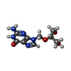
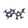
PDB-4d9h: Crystal structure of the hexameric purine nucleoside phosphorylase from Bacillus subtilis in complex with adenosine 手法: X-RAY DIFFRACTION / 解像度: 1.91 Å
PDB-4da0: Crystal structure of the hexameric purine nucleoside phosphorylase from Bacillus subtilis in complex with 2'-deoxyguanosine 手法: X-RAY DIFFRACTION / 解像度: 2.95 Å
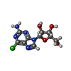
PDB-4da6: Crystal structure of the hexameric purine nucleoside phosphorylase from Bacillus subtilis in complex with ganciclovir 手法: X-RAY DIFFRACTION / 解像度: 1.7 Å
PDB-4da7: Crystal structure of the hexameric purine nucleoside phosphorylase from Bacillus subtilis in complex with aciclovir 手法: X-RAY DIFFRACTION / 解像度: 2.05 Å
PDB-4da8: Crystal structure of the hexameric purine nucleoside phosphorylase from Bacillus subtilis in complex with 8-bromoguanosine 手法: X-RAY DIFFRACTION / 解像度: 2.6 Å
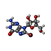
PDB-4dab: Crystal structure of the hexameric purine nucleoside phosphorylase from Bacillus subtilis in complex with hypoxanthine 手法: X-RAY DIFFRACTION / 解像度: 1.85 Å
PDB-4dae: Crystal structure of the hexameric purine nucleoside phosphorylase from Bacillus subtilis in complex with 6-chloroguanosine 手法: X-RAY DIFFRACTION / 解像度: 2.35 Å
PDB-4dan: Crystal structure of the hexameric purine nucleoside phosphorylase from Bacillus subtilis in complex with 2-fluoroadenosine 手法: X-RAY DIFFRACTION / 解像度: 2.56 Å
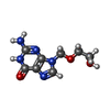
PDB-4dao: Crystal structure of the hexameric purine nucleoside phosphorylase from Bacillus subtilis in complex with adenine 手法: X-RAY DIFFRACTION / 解像度: 2.22 Å
PDB-4dar: Crystal structure of the hexameric purine nucleoside phosphorylase from Bacillus subtilis in complex with tubercidin 手法: X-RAY DIFFRACTION / 解像度: 3.15 Å
 ムービー
ムービー コントローラー
 構造ビューア
 万見文献について


 著者
著者 リンク
リンク


 キーワード
キーワード TRANSFERASE (転移酵素) / Phosphorylase/hydrolase-like
