+検索条件
-Structure paper
| タイトル | Zinc coordination geometry and ligand binding affinity: the structural and kinetic analysis of the second-shell serine 228 residue and the methionine 180 residue of the aminopeptidase from Vibrio proteolyticus. |
|---|---|
| ジャーナル・号・ページ | Biochemistry, Vol. 47, Page 7673-7683, Year 2008 |
| 掲載日 | 2007年10月19日 (構造データの登録日) |
 著者 著者 | Ataie, N.J. / Hoang, Q.Q. / Zahniser, M.P. / Tu, Y. / Milne, A. / Petsko, G.A. / Ringe, D. |
 リンク リンク |  Biochemistry / Biochemistry /  PubMed:18576673 PubMed:18576673 |
| 手法 | X線回折 |
| 解像度 | 1.1 - 1.75 Å |
| 構造データ |  PDB-3b35:  PDB-3b3c: 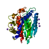 PDB-3b3s:  PDB-3b3t:  PDB-3b3v: 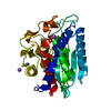 PDB-3b3w: 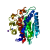 PDB-3b7i: |
| 化合物 |  ChemComp-ZN:  ChemComp-NA: 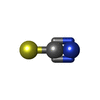 ChemComp-SCN:  ChemComp-HOH:  ChemComp-K:  ChemComp-PLU:  ChemComp-LEU:  ChemComp-ILE:  ChemComp-VAL: |
| 由来 |
|
 キーワード キーワード | HYDROLASE / alpha beta / Aminopeptidase / Metal-binding / Protease / Secreted / Zinc / Zymogen |
 ムービー
ムービー コントローラー
 構造ビューア
 万見文献について


 vibrio proteolyticus (バクテリア)