 ムービー
ムービー コントローラー
 構造ビューア
 万見文献について


+検索条件
-Structure paper
| タイトル | Substrate Recognition by the Multifunctional Cytochrome P450 Mycg in Mycinamicin Hydroxylation and Epoxidation Reactions. |
|---|---|
| ジャーナル・号・ページ | J. Biol. Chem., Vol. 287, Page 37880-, Year 2012 |
| 掲載日 | 2011年1月5日 (構造データの登録日) |
 著者 著者 | Li, S. / Tietz, D.R. / Rutaganira, F.U. / Kells, P.M. / Anzai, Y. / Kato, F. / Pochapsky, T.C. / Sherman, D.H. / Podust, L.M. |
 リンク リンク | J. Biol. Chem. / PubMed:22952225 |
| 手法 | X線回折 |
| 解像度 | 1.62 - 2.39 Å |
| 構造データ |  PDB-2y46: 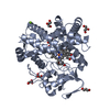 PDB-2y5n: 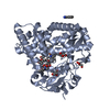 PDB-2y5z: 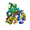 PDB-2y98:  PDB-2yca: 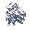 PDB-2ygx: 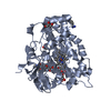 PDB-3zsn: 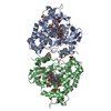 PDB-4aw3: |
| 化合物 |  ChemComp-HEM: 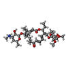 ChemComp-MIV:  ChemComp-BEN:  ChemComp-GOL:  ChemComp-HOH:  ChemComp-MYV:  ChemComp-MG: 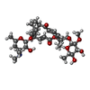 ChemComp-ZM3:  ChemComp-CL:  ChemComp-SO4:  ChemComp-SIN: |
| 由来 |
|
 キーワード キーワード | OXIDOREDUCTASE / MYCINAMICIN BIOSYNTHESIS / ELECTRON TRANSPORT / METAL BINDING |
 micromonospora griseorubida (バクテリア)