 ムービー
ムービー コントローラー
 構造ビューア
 万見文献について


+検索条件
-Structure paper
| タイトル | Structure of Ribosomal Silencing Factor Bound to Mycobacterium tuberculosis Ribosome. |
|---|---|
| ジャーナル・号・ページ | Structure, Vol. 23, Issue 10, Page 1858-1865, Year 2015 |
| 掲載日 | 2015年10月6日 |
 著者 著者 | Xiaojun Li / Qingan Sun / Cai Jiang / Kailu Yang / Li-Wei Hung / Junjie Zhang / James C Sacchettini /  |
| PubMed 要旨 | The ribosomal silencing factor RsfS slows cell growth by inhibiting protein synthesis during periods of diminished nutrient availability. The crystal structure of Mycobacterium tuberculosis (Mtb) ...The ribosomal silencing factor RsfS slows cell growth by inhibiting protein synthesis during periods of diminished nutrient availability. The crystal structure of Mycobacterium tuberculosis (Mtb) RsfS, together with the cryo-electron microscopy (EM) structure of the large subunit 50S of Mtb ribosome, reveals how inhibition of protein synthesis by RsfS occurs. RsfS binds to the 50S at L14, which, when occupied, blocks the association of the small subunit 30S. Although Mtb RsfS is a dimer in solution, only a single subunit binds to 50S. The overlap between the dimer interface and the L14 binding interface confirms that the RsfS dimer must first dissociate to a monomer in order to bind to L14. RsfS interacts primarily through electrostatic and hydrogen bonding to L14. The EM structure shows extended rRNA density that it is not found in the Escherichia coli ribosome, the most striking of these being the extended RNA helix of H54a. |
 リンク リンク | Structure / PubMed:26299947 / PubMed Central |
| 手法 | EM (単粒子) / X線回折 |
| 解像度 | 2.1 - 9.3 Å |
| 構造データ | 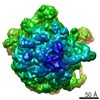 EMDB-6177:  EMDB-6178: 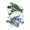 PDB-4wcw: |
| 化合物 |  ChemComp-MG:  ChemComp-MPD:  ChemComp-HOH: |
| 由来 |
|
 キーワード キーワード | TRANSLATION / Tuberculosis Ribosomal Silencing dimer / Structural Genomics / TB Structural Genomics Consortium / TBSGC |

 mycobacterium tuberculosis (結核菌)