 ムービー
ムービー コントローラー
 構造ビューア
 万見文献について


+検索条件
-Structure paper
| タイトル | Paired beta-sheet structure of an Abeta(1-40) amyloid fibril revealed by electron microscopy. |
|---|---|
| ジャーナル・号・ページ | Proc Natl Acad Sci U S A, Vol. 105, Issue 21, Page 7462-7466, Year 2008 |
| 掲載日 | 2008年5月27日 |
 著者 著者 | Carsten Sachse / Marcus Fändrich / Nikolaus Grigorieff /  |
| PubMed 要旨 | Alzheimer's disease is a neurodegenerative disorder that is characterized by the cerebral deposition of amyloid fibrils formed by Abeta peptide. Despite their prevalence in Alzheimer's and other ...Alzheimer's disease is a neurodegenerative disorder that is characterized by the cerebral deposition of amyloid fibrils formed by Abeta peptide. Despite their prevalence in Alzheimer's and other neurodegenerative diseases, important details of the structure of amyloid fibrils remain unknown. Here, we present a three-dimensional structure of a mature amyloid fibril formed by Abeta(1-40) peptide, determined by electron cryomicroscopy at approximately 8-A resolution. The fibril consists of two protofilaments, each containing approximately 5-nm-long regions of beta-sheet structure. A local twofold symmetry within each region suggests that pairs of beta-sheets are formed from equivalent parts of two Abeta(1-40) peptides contained in each protofilament. The pairing occurs via tightly packed interfaces, reminiscent of recently reported steric zipper structures. However, unlike these previous structures, the beta-sheet pairing is observed within an amyloid fibril and includes significantly longer amino acid sequences. |
 リンク リンク | Proc Natl Acad Sci U S A / PubMed:18483195 / PubMed Central |
| 手法 | EM (らせん対称) |
| 解像度 | 8.8 Å |
| 構造データ | 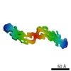 EMDB-5008: |
