 ムービー
ムービー コントローラー
 構造ビューア
 万見文献について


+検索条件
-Structure paper
| タイトル | Structure of myosin filaments from relaxed flight muscle by cryo-EM at 6 Å resolution. |
|---|---|
| ジャーナル・号・ページ | Sci Adv, Vol. 2, Issue 9, Page e1600058, Year 2016 |
| 掲載日 | 2016年9月30日 |
 著者 著者 | Zhongjun Hu / Dianne W Taylor / Michael K Reedy / Robert J Edwards / Kenneth A Taylor /  |
| PubMed 要旨 | We describe a cryo-electron microscopy three-dimensional image reconstruction of relaxed myosin II-containing thick filaments from the flight muscle of the giant water bug . The relaxed thick ...We describe a cryo-electron microscopy three-dimensional image reconstruction of relaxed myosin II-containing thick filaments from the flight muscle of the giant water bug . The relaxed thick filament structure is a key element of muscle physiology because it facilitates the reextension process following contraction. Conversely, the myosin heads must disrupt their relaxed arrangement to drive contraction. Previous models predicted that myosin was unique in having an intermolecular head-head interaction, as opposed to the intramolecular head-head interaction observed in all other species. In contrast to the predicted model, we find an intramolecular head-head interaction, which is similar to that of other thick filaments but oriented in a distinctly different way. The arrangement of myosin's long α-helical coiled-coil rod domain has been hypothesized as either curved layers or helical subfilaments. Our reconstruction is the first report having sufficient resolution to track the rod α helices in their native environment at resolutions ~5.5 Å, and it shows that the layer arrangement is correct for . Threading separate paths through the forest of myosin coiled coils are four nonmyosin peptides. We suggest that the unusual position of the heads and the rod arrangement separated by nonmyosin peptides are adaptations for mechanical signal transduction whereby applied tension disrupts the myosin heads as a component of stretch activation. |
 リンク リンク | Sci Adv / PubMed:27704041 / PubMed Central |
| 手法 | EM (単粒子) |
| 解像度 | 5.5 Å |
| 構造データ | 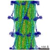 EMDB-3301: |
| 由来 |
|
 Lethocerus indicus (昆虫)