ムービー
ムービー コントローラー
 構造ビューア
 万見文献について


+検索条件
-Structure paper
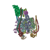
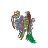
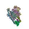
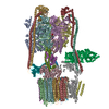
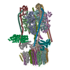
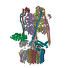
| タイトル | Coordinated conformational changes in the V complex during V-ATPase reversible dissociation. |
|---|---|
| ジャーナル・号・ページ | Nat Struct Mol Biol, Vol. 29, Issue 5, Page 430-439, Year 2022 |
| 掲載日 | 2022年4月25日 |
 著者 著者 | Thamiya Vasanthakumar / Kristine A Keon / Stephanie A Bueler / Michael C Jaskolka / John L Rubinstein /   |
| PubMed 要旨 | Vacuolar-type ATPases (V-ATPases) are rotary enzymes that acidify intracellular compartments in eukaryotic cells. These multi-subunit complexes consist of a cytoplasmic V region that hydrolyzes ATP ...Vacuolar-type ATPases (V-ATPases) are rotary enzymes that acidify intracellular compartments in eukaryotic cells. These multi-subunit complexes consist of a cytoplasmic V region that hydrolyzes ATP and a membrane-embedded V region that transports protons. V-ATPase activity is regulated by reversible dissociation of the two regions, with the isolated V and V complexes becoming autoinhibited on disassembly and subunit C subsequently detaching from V. In yeast, assembly of the V and V regions is mediated by the regulator of the ATPase of vacuoles and endosomes (RAVE) complex through an unknown mechanism. We used cryogenic-electron microscopy of yeast V-ATPase to determine structures of the intact enzyme, the dissociated but complete V complex and the V complex lacking subunit C. On separation, V undergoes a dramatic conformational rearrangement, with its rotational state becoming incompatible for reassembly with V. Loss of subunit C allows V to match the rotational state of V, suggesting how RAVE could reassemble V and V by recruiting subunit C. |
 リンク リンク | Nat Struct Mol Biol / PubMed:35469063 |
| 手法 | EM (単粒子) |
| 解像度 | 3.3 - 3.8 Å |
| 構造データ | EMDB-25996, PDB-7tmm: EMDB-25997, PDB-7tmo: EMDB-25998, PDB-7tmp: EMDB-25999, PDB-7tmq: EMDB-26000, PDB-7tmr: EMDB-26001, PDB-7tms: EMDB-26002, PDB-7tmt: |
| 化合物 |  ChemComp-ADP:  ChemComp-ATP:  ChemComp-MG: |
| 由来 |
|
 キーワード キーワード | HYDROLASE / V-ATPase / MEMBRANE PROTEIN |