 ムービー
ムービー コントローラー
 構造ビューア
 万見文献について


+検索条件
-Structure paper
| タイトル | Three-dimensional reconstruction of Agrobacterium VirE2 protein with single-stranded DNA. |
|---|---|
| ジャーナル・号・ページ | J Biol Chem, Vol. 279, Issue 24, Page 25359-25363, Year 2004 |
| 掲載日 | 2004年6月11日 |
 著者 著者 | Asmahan Abu-Arish / Daphna Frenkiel-Krispin / Tobin Fricke / Tzvi Tzfira / Vitaly Citovsky / Sharon Grayer Wolf / Michael Elbaum /  |
| PubMed 要旨 | Agrobacterium tumefaciens infects plant cells by a unique mechanism involving an interkingdom genetic transfer. A single-stranded DNA substrate is transported across the two cell walls along with the ...Agrobacterium tumefaciens infects plant cells by a unique mechanism involving an interkingdom genetic transfer. A single-stranded DNA substrate is transported across the two cell walls along with the bacterial virulence proteins VirD2 and VirE2. A single VirD2 molecule covalently binds to the 5'-end of the single-stranded DNA, while the VirE2 protein binds stoichiometrically along the length of the DNA, without sequence specificity. An earlier transmission/scanning transmission electron microscopy study indicated a solenoidal ("telephone coil") organization of the VirE2-DNA complex. Here we report a three-dimensional reconstruction of this complex using electron microscopy and single-particle image-processing methods. We find a hollow helical structure of 15.7-nm outer diameter, with a helical rise of 51.5 nm and 4.25 VirE2 proteins/turn. The inner face of the protein units contains a continuous wall and an inward protruding shelf. These structures appear to accommodate the DNA binding. Such a quaternary arrangement naturally sequesters the DNA from cytoplasmic nucleases and suggests a mechanism for its nuclear import by decoration with host cell factors. Coexisting with the helices, we also found VirE2 tetrameric ring structures. A two-dimensional average of the latter confirms the major features of the three-dimensional reconstruction. |
 リンク リンク | J Biol Chem / PubMed:15054095 |
| 手法 | EM (らせん対称) |
| 解像度 | 25.0 Å |
| 構造データ | 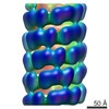 EMDB-2368: |
| 由来 |
|
 Agrobacterium fabrum str. C58 (バクテリア)