 ムービー
ムービー コントローラー
 構造ビューア
 万見文献について


+検索条件
-Structure paper
| タイトル | Family-wide Structural and Biophysical Analysis of Binding Interactions among Non-clustered δ-Protocadherins. |
|---|---|
| ジャーナル・号・ページ | Cell Rep, Vol. 30, Issue 8, Page 2655-22671.e7, Year 2020 |
| 掲載日 | 2020年2月25日 |
 著者 著者 | Oliver J Harrison / Julia Brasch / Phinikoula S Katsamba / Goran Ahlsen / Alex J Noble / Hanbin Dan / Rosemary V Sampogna / Clinton S Potter / Bridget Carragher / Barry Honig / Lawrence Shapiro /  |
| PubMed 要旨 | Non-clustered δ1- and δ2-protocadherins, close relatives of clustered protocadherins, function in cell adhesion and motility and play essential roles in neural patterning. To understand the ...Non-clustered δ1- and δ2-protocadherins, close relatives of clustered protocadherins, function in cell adhesion and motility and play essential roles in neural patterning. To understand the molecular interactions underlying these functions, we used solution biophysics to characterize binding of δ1- and δ2-protocadherins, determined crystal structures of ectodomain complexes from each family, and assessed ectodomain assembly in reconstituted intermembrane junctions by cryoelectron tomography (cryo-ET). Homophilic trans (cell-cell) interactions were preferred for all δ-protocadherins, with additional weaker heterophilic interactions observed exclusively within each subfamily. As expected, δ1- and δ2-protocadherin trans dimers formed through antiparallel EC1-EC4 interfaces, like clustered protocadherins. However, no ectodomain-mediated cis (same-cell) interactions were detectable in solution; consistent with this, cryo-ET of reconstituted junctions revealed dense assemblies lacking the characteristic order observed for clustered protocadherins. Our results define non-clustered protocadherin binding properties and their structural basis, providing a foundation for interpreting their functional roles in neural patterning. |
 リンク リンク | Cell Rep / PubMed:32101743 / PubMed Central |
| 手法 | EM (トモグラフィー) / X線回折 |
| 解像度 | 2 - 3.71 Å |
| 構造データ |  EMDB-21188:  EMDB-21189:  EMDB-21190:  EMDB-21191:  EMDB-21192:  EMDB-21193: 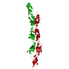 PDB-6vfp:  PDB-6vfq:  PDB-6vfr: 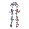 PDB-6vft:  PDB-6vfu:  PDB-6vfv:  PDB-6vfw:  PDB-6vg1:  PDB-6vg4: |
| 化合物 |  ChemComp-NAG:  ChemComp-CA:  ChemComp-HOH:  ChemComp-MAN:  ChemComp-ACT:  ChemComp-EDO:  ChemComp-NA:  ChemComp-NI:  ChemComp-CL:  ChemComp-TAM: |
| 由来 |
|
 キーワード キーワード | CELL ADHESION / cadherin extracellular region / non-clustered delta1 family protocadherin / homophilic adhesion/recognition calcium-dependent adhesion molecule / non-clustered delta2 family protocadherin / non-clustered delta2 family / protocadherin homophilic adhesion/recognition calcium-dependent / adhesion molecule / protocadherin / homophilic / adhesion/recognition calcium-dependent adhesion molecule / non-clustered delta2 protocadherin / homophilic adhesion/recognition calcium-dependent / cadherin / hemophilic / non-clustered / calcium binding |
 homo sapiens (ヒト)