 ムービー
ムービー コントローラー
 構造ビューア
 万見文献について


+検索条件
-Structure paper
| タイトル | 3D Ultrastructure of the Cochlear Outer Hair Cell Lateral Wall Revealed By Electron Tomography. |
|---|---|
| ジャーナル・号・ページ | Front Cell Neurosci, Vol. 13, Page 560, Year 2019 |
| 掲載日 | 2019年12月20日 |
 著者 著者 | William Jeffrey Triffo / Hildur Palsdottir / Junha Song / David Gene Morgan / Kent L McDonald / Manfred Auer / Robert M Raphael /  |
| PubMed 要旨 | Outer Hair Cells (OHCs) in the mammalian cochlea display a unique type of voltage-induced mechanical movement termed electromotility, which amplifies auditory signals and contributes to the ...Outer Hair Cells (OHCs) in the mammalian cochlea display a unique type of voltage-induced mechanical movement termed electromotility, which amplifies auditory signals and contributes to the sensitivity and frequency selectivity of mammalian hearing. Electromotility occurs in the OHC lateral wall, but it is not fully understood how the supramolecular architecture of the lateral wall enables this unique form of cellular motility. Employing electron tomography of high-pressure frozen and freeze-substituted OHCs, we visualized the 3D structure and organization of the membrane and cytoskeletal components of the OHC lateral wall. The subsurface cisterna (SSC) is a highly prominent feature, and we report that the SSC membranes and lumen possess hexagonally ordered arrays of particles. We also find the SSC is tightly connected to adjacent actin filaments by short filamentous protein connections. Pillar proteins that join the plasma membrane to the cytoskeleton appear as variable structures considerably thinner than actin filaments and significantly more flexible than actin-SSC links. The structurally rich organization and rigidity of the SSC coupled with apparently weaker mechanical connections between the plasma membrane (PM) and cytoskeleton reveal that the membrane-cytoskeletal architecture of the OHC lateral wall is more complex than previously appreciated. These observations are important for our understanding of OHC mechanics and need to be considered in computational models of OHC electromotility that incorporate subcellular features. |
 リンク リンク | Front Cell Neurosci / PubMed:31920560 / PubMed Central |
| 手法 | EM (トモグラフィー) |
| 構造データ | 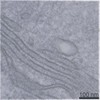 EMDB-20547: |
| 由来 |
|
 Cavia porcellus (哺乳類)