 ムービー
ムービー コントローラー
 構造ビューア
 万見文献について


+検索条件
-Structure paper
| タイトル | Cryo electron tomography of herpes simplex virus during axonal transport and secondary envelopment in primary neurons. |
|---|---|
| ジャーナル・号・ページ | PLoS Pathog, Vol. 7, Issue 12, Page e1002406, Year 2011 |
| 掲載日 | 2011年12月15日 |
 著者 著者 | Iosune Ibiricu / Juha T Huiskonen / Katinka Döhner / Frank Bradke / Beate Sodeik / Kay Grünewald /  |
| PubMed 要旨 | During herpes simplex virus 1 (HSV1) egress in neurons, viral particles travel from the neuronal cell body along the axon towards the synapse. Whether HSV1 particles are transported as enveloped ...During herpes simplex virus 1 (HSV1) egress in neurons, viral particles travel from the neuronal cell body along the axon towards the synapse. Whether HSV1 particles are transported as enveloped virions as proposed by the 'married' model or as non-enveloped capsids suggested by the 'separate' model is controversial. Specific viral proteins may form a recruitment platform for microtubule motors that catalyze such transport. However, their subviral location has remained elusive. Here we established a system to analyze herpesvirus egress by cryo electron tomography. At 16 h post infection, we observed intra-axonal transport of progeny HSV1 viral particles in dissociated hippocampal neurons by live-cell fluorescence microscopy. Cryo electron tomography of frozen-hydrated neurons revealed that most egressing capsids were transported independently of the viral envelope. Unexpectedly, we found not only DNA-containing capsids (cytosolic C-capsids), but also capsids lacking DNA (cytosolic A-/B-capsids) in mid-axon regions. Subvolume averaging revealed lower amounts of tegument on cytosolic A-/B-capsids than on C-capsids. Nevertheless, all capsid types underwent active axonal transport. Therefore, even few tegument proteins on the capsid vertices seemed to suffice for transport. Secondary envelopment of capsids was observed at axon terminals. On their luminal face, the enveloping vesicles were studded with typical glycoprotein-like spikes. Furthermore, we noted an accretion of tegument density at the concave cytosolic face of the vesicle membrane in close proximity to the capsids. Three-dimensional analysis revealed that these assembly sites lacked cytoskeletal elements, but that filamentous actin surrounded them and formed an assembly compartment. Our data support the 'separate model' for HSV1 egress, i.e. progeny herpes viruses being transported along axons as subassemblies and not as complete virions within transport vesicles. |
 リンク リンク | PLoS Pathog / PubMed:22194682 / PubMed Central |
| 手法 | EM (サブトモグラム平均) |
| 解像度 | 56.0 - 97.0 Å |
| 構造データ | 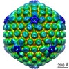 EMDB-1956: 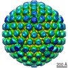 EMDB-1957: 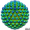 EMDB-1958:  EMDB-1959: |
