 ムービー
ムービー コントローラー
 構造ビューア
 万見文献について


+検索条件
-Structure paper
| タイトル | Cryo-electron microscopy study of bacteriophage T4 displaying anthrax toxin proteins. |
|---|---|
| ジャーナル・号・ページ | Virology, Vol. 367, Issue 2, Page 422-427, Year 2007 |
| 掲載日 | 2007年10月25日 |
 著者 著者 | Andrei Fokine / Valorie D Bowman / Anthony J Battisti / Qin Li / Paul R Chipman / Venigalla B Rao / Michael G Rossmann /  |
| PubMed 要旨 | The bacteriophage T4 capsid contains two accessory surface proteins, the small outer capsid protein (Soc, 870 copies) and the highly antigenic outer capsid protein (Hoc, 155 copies). As these are ...The bacteriophage T4 capsid contains two accessory surface proteins, the small outer capsid protein (Soc, 870 copies) and the highly antigenic outer capsid protein (Hoc, 155 copies). As these are dispensable for capsid formation, they can be used for displaying proteins and macromolecular complexes on the T4 capsid surface. Anthrax toxin components were attached to the T4 capsid as a fusion protein of the N-terminal domain of the anthrax lethal factor (LFn) with Soc. The LFn-Soc fusion protein was complexed in vitro with Hoc(-)Soc(-)T4 phage. Subsequently, cleaved anthrax protective antigen heptamers (PA63)(7) were attached to the exposed LFn domains. A cryo-electron microscopy study of the decorated T4 particles shows the complex of PA63 heptamers with LFn-Soc on the phage surface. Although the cryo-electron microscopy reconstruction is unable to differentiate on its own between different proposed models of the anthrax toxin, the density is consistent with a model that had predicted the orientation and position of three LFn molecules bound to one PA63 heptamer. |
 リンク リンク | Virology / PubMed:17624389 / PubMed Central |
| 手法 | EM (単粒子) |
| 解像度 | 35.0 Å |
| 構造データ | 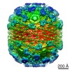 EMDB-1280: |
| 由来 |
|

