 ムービー
ムービー コントローラー
 構造ビューア
 万見文献について


+検索条件
-Structure paper
| タイトル | Structural basis for the interaction of protein S1 with the Escherichia coli ribosome. |
|---|---|
| ジャーナル・号・ページ | Nucleic Acids Res, Vol. 43, Issue 1, Page 661-673, Year 2015 |
| 掲載日 | 2014年12月15日 |
 著者 著者 | Konstantin Byrgazov / Irina Grishkovskaya / Stefan Arenz / Nicolas Coudevylle / Hannes Temmel / Daniel N Wilson / Kristina Djinovic-Carugo / Isabella Moll /    |
| PubMed 要旨 | In Gram-negative bacteria, the multi-domain protein S1 is essential for translation initiation, as it recruits the mRNA and facilitates its localization in the decoding centre. In sharp contrast to ...In Gram-negative bacteria, the multi-domain protein S1 is essential for translation initiation, as it recruits the mRNA and facilitates its localization in the decoding centre. In sharp contrast to its functional importance, S1 is still lacking from the high-resolution structures available for Escherichia coli and Thermus thermophilus ribosomes and thus the molecular mechanism governing the S1-ribosome interaction has still remained elusive. Here, we present the structure of the N-terminal S1 domain D1 when bound to the ribosome at atomic resolution by using a combination of NMR, X-ray crystallography and cryo-electron microscopy. Together with biochemical assays, the structure reveals that S1 is anchored to the ribosome primarily via a stabilizing π-stacking interaction within the short but conserved N-terminal segment that is flexibly connected to domain D1. This interaction is further stabilized by salt bridges involving the zinc binding pocket of protein S2. Overall, this work provides one hitherto enigmatic piece in the 'ribosome puzzle', namely the detailed molecular insight into the topology of the S1-ribosome interface. Moreover, our data suggest novel mechanisms that have the potential to modulate protein synthesis in response to environmental cues by changing the affinity of S1 for the ribosome. |
 リンク リンク | Nucleic Acids Res / PubMed:25510494 / PubMed Central |
| 手法 | EM (単粒子) / X線回折 |
| 解像度 | 2.3 - 9.5 Å |
| 構造データ | 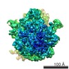 EMDB-6211: 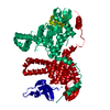 PDB-4toi: |
| 化合物 |  ChemComp-ZN:  ChemComp-HOH: |
| 由来 |
|
 キーワード キーワード |  RIBOSOMAL PROTEIN / complex / translation (翻訳 (生物学)) RIBOSOMAL PROTEIN / complex / translation (翻訳 (生物学)) |

