Jinghua Tang / Junhua Pan / Wendy M Havens / Wendy F Ochoa / Tom S Y Guu / Said A Ghabrial / Max L Nibert / Yizhi Jane Tao / Timothy S Baker /
PubMed 要旨
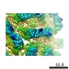
Most dsRNA viruses have a genome-enclosing capsid that comprises 120 copies of a single coat protein (CP). These 120 CP subunits are arranged as asymmetrical dimers that surround the icosahedral ...Most dsRNA viruses have a genome-enclosing capsid that comprises 120 copies of a single coat protein (CP). These 120 CP subunits are arranged as asymmetrical dimers that surround the icosahedral fivefold axes, forming pentamers of dimers that are thought to be assembly intermediates. This scheme is violated, however, in recent structures of two dsRNA viruses, a fungal virus from family Partitiviridae and a rabbit virus from family Picobirnaviridae, both of which have 120 CP subunits organized as dimers of quasisymmetrical dimers. In this study, we report the CP backbone trace of a second fungal partitivirus, determined in this case by electron cryomicroscopy and homology modeling. This virus also exhibits quasisymmetrical CP dimers that are connected by prominent surface arches and stabilized by domain swapping between the two CP subunits. The CP fold is dominated by alpha-helices, although beta-strands mediate several important contacts. A dimer-of-dimers assembly intermediate is again implicated. The disordered N-terminal tail of each CP subunit protrudes into the particle interior and likely interacts with the genome during packaging and/or transcription. These results broaden our understanding of conserved and variable aspects of partitivirus structure and reflect the growing use of electron cryomicroscopy for atomic modeling of protein folds.
EMDB-5161: PsV-F PDB-3iym: Backbone Trace of the Capsid Protein Dimer of a Fungal Partitivirus from Electron Cryomicroscopy and Homology Modeling 手法: EM (単粒子)
EMDB-5163: PsV-S PDB-3iym: Backbone Trace of the Capsid Protein Dimer of a Fungal Partitivirus from Electron Cryomicroscopy and Homology Modeling 手法: EM (単粒子)
 ムービー
ムービー コントローラー
 構造ビューア
 万見文献について


 著者
著者
 リンク
リンク キーワード
キーワード VIRUS (ウイルス) /
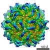

 penicillium stoloniferum virus s (ウイルス)