ムービー
ムービー コントローラー
 構造ビューア
 EMN文献について


+検索条件
-Structure paper
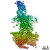
| タイトル | Structural insights into differences in G protein activation by family A and family B GPCRs. |
|---|---|
| ジャーナル・号・ページ | Science, Vol. 369, Issue 6503, Year 2020 |
| 掲載日 | 2020年7月31日 |
 著者 著者 | Daniel Hilger / Kaavya Krishna Kumar / Hongli Hu / Mie Fabricius Pedersen / Evan S O'Brien / Lise Giehm / Christine Jennings / Gözde Eskici / Asuka Inoue / Michael Lerch / Jesper Mosolff Mathiesen / Georgios Skiniotis / Brian K Kobilka /    |
| PubMed 要旨 | Family B heterotrimeric guanine nucleotide-binding protein (G protein)-coupled receptors (GPCRs) play important roles in carbohydrate metabolism. Recent structures of family B GPCR-G protein ...Family B heterotrimeric guanine nucleotide-binding protein (G protein)-coupled receptors (GPCRs) play important roles in carbohydrate metabolism. Recent structures of family B GPCR-G protein complexes reveal a disruption in the α-helix of transmembrane segment 6 (TM6) not observed in family A GPCRs. To investigate the functional impact of this structural difference, we compared the structure and function of the glucagon receptor (GCGR; family B) with the β adrenergic receptor (βAR; family A). We determined the structure of the GCGR-G complex by means of cryo-electron microscopy at 3.1-angstrom resolution. This structure shows the distinct break in TM6. Guanosine triphosphate (GTP) turnover, guanosine diphosphate release, GTP binding, and G protein dissociation studies revealed much slower rates for G protein activation by the GCGR compared with the βAR. Fluorescence and double electron-electron resonance studies suggest that this difference is due to the inability of agonist alone to induce a detectable outward movement of the cytoplasmic end of TM6. |
 リンク リンク | Science / PubMed:32732395 / PubMed Central |
| 手法 | EM (単粒子) |
| 解像度 | 3.1 Å |
| 構造データ | EMDB-21866, PDB-6wpw: |
| 由来 |
|
 キーワード キーワード |  SIGNALING PROTEIN/HORMONE/IMMUNE SYSTEM / GPCR (Gタンパク質共役受容体) / G protein (Gタンパク質) / glucagon (グルカゴン) / signaling / complex / SIGNALING PROTEIN-HORMONE-IMMUNE SYSTEM complex SIGNALING PROTEIN/HORMONE/IMMUNE SYSTEM / GPCR (Gタンパク質共役受容体) / G protein (Gタンパク質) / glucagon (グルカゴン) / signaling / complex / SIGNALING PROTEIN-HORMONE-IMMUNE SYSTEM complex |