[English] 日本語
Yorodumi Papers
- Database of articles cited by EMDB/PDB/SASBDB data -

+
Search query

Keywords
Structure methods
Author
Journal
IF

-
Structure paper

TitleStructural comparison of different antibodies interacting with parvovirus capsids.
Journal, issue, pagesJ Virol, Vol. 83, Issue 11, Page 5556-5566, Year 2009
Publish dateMar 25, 2009
AuthorsSusan Hafenstein / Valorie D Bowman / Tao Sun / Christian D S Nelson / Laura M Palermo / Paul R Chipman / Anthony J Battisti / Colin R Parrish / Michael G Rossmann /
PubMed AbstractThe structures of canine parvovirus (CPV) and feline parvovirus (FPV) complexed with antibody fragments from eight different neutralizing monoclonal antibodies were determined by cryo-electron ...The structures of canine parvovirus (CPV) and feline parvovirus (FPV) complexed with antibody fragments from eight different neutralizing monoclonal antibodies were determined by cryo-electron microscopy (cryoEM) reconstruction to resolutions varying from 8.5 to 18 A. The crystal structure of one of the Fab molecules and the sequence of the variable domain for each of the Fab molecules have been determined. The structures of Fab fragments not determined crystallographically were predicted by homology modeling according to the amino acid sequence. Fitting of the Fab and virus structures into the cryoEM densities identified the footprints of each antibody on the viral surface. As anticipated from earlier analyses, the Fab binding sites are directed to two epitopes, A and B. The A site is on an exposed part of the surface near an icosahedral threefold axis, whereas the B site is about equidistant from the surrounding five-, three-, and twofold axes. One antibody directed to the A site binds CPV but not FPV. Two of the antibodies directed to the B site neutralize the virus as Fab fragments. The differences in antibody properties have been linked to the amino acids within the antibody footprints, the position of the binding site relative to the icosahedral symmetry elements, and the orientation of the Fab structure relative to the surface of the virus. Most of the exposed surface area was antigenic, although each of the antibodies had a common area of overlap that coincided with the positions of the previously mapped escape mutations.
External linksJ Virol / PubMed:19321620 / PubMed Central
MethodsEM (single particle) / X-ray diffraction
Resolution2 - 18.0 Å
Structure data

EMDB-5105: Canine parvovirus in complex with FAb from neutralizing antibody MAb 14
PDB-3iy0: Variable domains of the x-ray structure of Fab 14 fitted into the cryoEM reconstruction of the virus-Fab 14 complex
Method: EM (single particle) / Resolution: 12.5 Å

EMDB-5106: Fab from MAb B interacting with feline panleukopenia virus
PDB-3iy1: Variable domains of the WAM of Fab B fitted into the cryoEM reconstruction of the virus-Fab B complex
Method: EM (single particle) / Resolution: 18.0 Å

EMDB-5107: Feline panleukopenia virus in complex with FAb from neutralizing antibody MAb 6
PDB-3iy2: Variable domains of the computer generated model (WAM) of Fab 6 fitted into the cryoEM reconstruction of the virus-Fab 6 complex
Method: EM (single particle) / Resolution: 18.0 Å

EMDB-5108: Feline panleukopenia virus in complex with FAb from neutralizing antibody MAb 8
PDB-3iy3: Variable domains of the computer generated model (WAM) of Fab 8 fitted into the cryoEM reconstruction of the virus-Fab 8 complex
Method: EM (single particle) / Resolution: 11.1 Å

EMDB-5109: Feline panleukopenia virus in complex with FAb from neutralizing antibody MAb 15
PDB-3iy4: Variable domains of the computer generated model (WAM) of Fab 15 fitted into the cryoEM reconstruction of the virus-Fab 15 complex
Method: EM (single particle) / Resolution: 11.7 Å

EMDB-5110: Feline panleukopenia virus in complex with FAb from neutralizing antibody MAb 16
PDB-3iy5: Variable domains of the mouse Fab (1AIF) fitted into the cryoEM reconstruction of the virus-Fab 16 complex
Method: EM (single particle) / Resolution: 18.0 Å

EMDB-5111: Feline panleukopenia virus in complex with FAb from neutralizing antibody MAb E
PDB-3iy6: Variable domains of the computer generated model (WAM) of Fab E fitted into the cryoEM reconstruction of the virus-Fab E complex
Method: EM (single particle) / Resolution: 12.0 Å

EMDB-5112: Feline panleukopenia virus in complex with FAb from neutralizing antibody MAb F
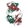
PDB-3iy7: Variable domains of the computer generated model (WAM) of Fab F fitted into the cryoEM reconstruction of the virus-Fab F complex
Method: EM (single particle) / Resolution: 14.0 Å

PDB-3gk8:
X-ray crystal structure of the Fab from MAb 14, mouse antibody against Canine Parvovirus
Method: X-RAY DIFFRACTION / Resolution: 2 Å

Chemicals

ChemComp-HOH:
WATER

Source
  • mus musculus (house mouse)
  • rattus norvegicus (Norway rat)
KeywordsIMMUNE SYSTEM / antibody fragment from neutralizing monoclonal antibody against canine parvovirus / cryoEM / neutralizing antibody / parvovirus / canine / feline / fab footprint

+
About Yorodumi Papers

-
News

-
Feb 9, 2022. New format data for meta-information of EMDB entries

New format data for meta-information of EMDB entries

  • Version 3 of the EMDB header file is now the official format.
  • The previous official version 1.9 will be removed from the archive.

Related info.:EMDB header

External links:wwPDB to switch to version 3 of the EMDB data model

-
Aug 12, 2020. Covid-19 info

Covid-19 info

URL: https://pdbj.org/emnavi/covid19.php

New page: Covid-19 featured information page in EM Navigator.

Related info.:Covid-19 info / Mar 5, 2020. Novel coronavirus structure data

+
Mar 5, 2020. Novel coronavirus structure data

Novel coronavirus structure data

Related info.:Yorodumi Speices / Aug 12, 2020. Covid-19 info

External links:COVID-19 featured content - PDBj / Molecule of the Month (242):Coronavirus Proteases

+
Jan 31, 2019. EMDB accession codes are about to change! (news from PDBe EMDB page)

EMDB accession codes are about to change! (news from PDBe EMDB page)

  • The allocation of 4 digits for EMDB accession codes will soon come to an end. Whilst these codes will remain in use, new EMDB accession codes will include an additional digit and will expand incrementally as the available range of codes is exhausted. The current 4-digit format prefixed with “EMD-” (i.e. EMD-XXXX) will advance to a 5-digit format (i.e. EMD-XXXXX), and so on. It is currently estimated that the 4-digit codes will be depleted around Spring 2019, at which point the 5-digit format will come into force.
  • The EM Navigator/Yorodumi systems omit the EMD- prefix.

Related info.:Q: What is EMD? / ID/Accession-code notation in Yorodumi/EM Navigator

External links:EMDB Accession Codes are Changing Soon! / Contact to PDBj

+
Jul 12, 2017. Major update of PDB

Major update of PDB

  • wwPDB released updated PDB data conforming to the new PDBx/mmCIF dictionary.
  • This is a major update changing the version number from 4 to 5, and with Remediation, in which all the entries are updated.
  • In this update, many items about electron microscopy experimental information are reorganized (e.g. em_software).
  • Now, EM Navigator and Yorodumi are based on the updated data.

External links:wwPDB Remediation / Enriched Model Files Conforming to OneDep Data Standards Now Available in the PDB FTP Archive

-
Yorodumi Papers

Database of articles cited by EMDB/PDB/SASBDB data

  • Database of articles cited by EMDB, PDB, and SASBDB entries
  • Using PubMed data

Related info.:EMDB / PDB / SASBDB / Yorodumi / EMN Papers / Changes in new EM Navigator and Yorodumi

Read more