 Movie
Movie Controller

[English] 日本語
 Yorodumi
- EMDB-5109: Feline panleukopenia virus in complex with FAb from neutralizing ... -
+ Open data

- Basic information
| Entry | Database: EMDB / ID: EMD-5109 | |||||||||
|---|---|---|---|---|---|---|---|---|---|---|
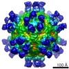
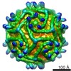
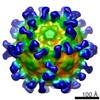
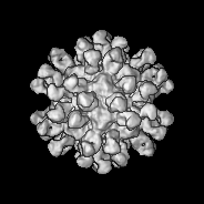
| Title | Feline panleukopenia virus in complex with FAb from neutralizing antibody MAb 15 | |||||||||
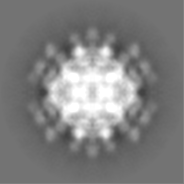
 Map data Map data | This is a map of the Fab from MAb 8 interacting with feline panleukopenia virus | |||||||||
 Sample Sample |
| |||||||||
 Keywords Keywords | parvovirus / antigenic epitope / antibody / Fab / neutralizing | |||||||||
| Biological species |  Feline panleukopenia virus Feline panleukopenia virus | |||||||||
| Method | single particle reconstruction / cryo EM / Resolution: 11.7 Å | |||||||||
 Authors Authors | Hafenstein S / Bowman VD / Sun T / Nelson CDS / Palermo LM / Chipman PR / Battisti AJ / Parrish CR / Rossmann MG | |||||||||
 Citation Citation | Journal: J Virol / Year: 2009 Title: Structural comparison of different antibodies interacting with parvovirus capsids. Authors: Susan Hafenstein / Valorie D Bowman / Tao Sun / Christian D S Nelson / Laura M Palermo / Paul R Chipman / Anthony J Battisti / Colin R Parrish / Michael G Rossmann /  Abstract: The structures of canine parvovirus (CPV) and feline parvovirus (FPV) complexed with antibody fragments from eight different neutralizing monoclonal antibodies were determined by cryo-electron ...The structures of canine parvovirus (CPV) and feline parvovirus (FPV) complexed with antibody fragments from eight different neutralizing monoclonal antibodies were determined by cryo-electron microscopy (cryoEM) reconstruction to resolutions varying from 8.5 to 18 A. The crystal structure of one of the Fab molecules and the sequence of the variable domain for each of the Fab molecules have been determined. The structures of Fab fragments not determined crystallographically were predicted by homology modeling according to the amino acid sequence. Fitting of the Fab and virus structures into the cryoEM densities identified the footprints of each antibody on the viral surface. As anticipated from earlier analyses, the Fab binding sites are directed to two epitopes, A and B. The A site is on an exposed part of the surface near an icosahedral threefold axis, whereas the B site is about equidistant from the surrounding five-, three-, and twofold axes. One antibody directed to the A site binds CPV but not FPV. Two of the antibodies directed to the B site neutralize the virus as Fab fragments. The differences in antibody properties have been linked to the amino acids within the antibody footprints, the position of the binding site relative to the icosahedral symmetry elements, and the orientation of the Fab structure relative to the surface of the virus. Most of the exposed surface area was antigenic, although each of the antibodies had a common area of overlap that coincided with the positions of the previously mapped escape mutations. | |||||||||
| History |
|
- Structure visualization
| Movie |
 Movie viewer Movie viewer |
|---|---|
| Structure viewer | EM map: SurfViewMolmilJmol/JSmol |
| Supplemental images |
 UCSF Chimera
- Downloads & links
-EMDB archive
| Map data | emd_5109.map.gz | 3.6 MB | EMDB map data format | |
|---|---|---|---|---|
| Header (meta data) | emd-5109-v30.xmlemd-5109.xml | 10.3 KB 10.3 KB | Display Display | EMDB header |
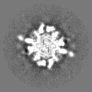
| Images | 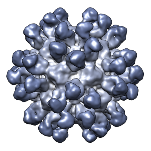 emd_5109_1.png emd_5109_1.png | 247.5 KB | ||
| Archive directory |  http://ftp.pdbj.org/pub/emdb/structures/EMD-5109ftp://ftp.pdbj.org/pub/emdb/structures/EMD-5109 http://ftp.pdbj.org/pub/emdb/structures/EMD-5109ftp://ftp.pdbj.org/pub/emdb/structures/EMD-5109 | HTTPS FTP |
-Related structure data
| Related structure data | 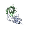 3iy4MC 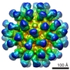 5105C 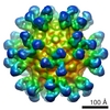 5106C 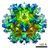 5107C 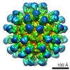 5108C 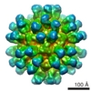 5110C 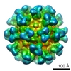 5111C 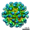 5112C 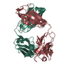 3gk8C 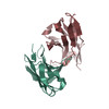 3iy0C 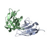 3iy1C 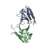 3iy2C  3iy3C 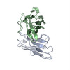 3iy5C 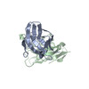 3iy6C 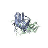 3iy7C M: atomic model generated by this map C: citing same article ( |
|---|---|
| Similar structure data |
-Links
| EMDB pages | EMDB (EBI/PDBe) / EMDataResource |
|---|
-Map
| File | Download / File: emd_5109.map.gz / Format: CCP4 / Size: 23.2 MB / Type: IMAGE STORED AS FLOATING POINT NUMBER (4 BYTES) | ||||||||||||||||||||||||||||||||||||||||||||||||||||||||||||||||||||
|---|---|---|---|---|---|---|---|---|---|---|---|---|---|---|---|---|---|---|---|---|---|---|---|---|---|---|---|---|---|---|---|---|---|---|---|---|---|---|---|---|---|---|---|---|---|---|---|---|---|---|---|---|---|---|---|---|---|---|---|---|---|---|---|---|---|---|---|---|---|
| Annotation | This is a map of the Fab from MAb 8 interacting with feline panleukopenia virus | ||||||||||||||||||||||||||||||||||||||||||||||||||||||||||||||||||||
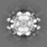
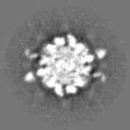
| Projections & slices | Image control
Images are generated by Spider. | ||||||||||||||||||||||||||||||||||||||||||||||||||||||||||||||||||||
| Voxel size | X=Y=Z: 2.87 Å | ||||||||||||||||||||||||||||||||||||||||||||||||||||||||||||||||||||
| Density |
| ||||||||||||||||||||||||||||||||||||||||||||||||||||||||||||||||||||
| Symmetry | Space group: 1 | ||||||||||||||||||||||||||||||||||||||||||||||||||||||||||||||||||||
| Details | EMDB XML:
CCP4 map header:
| ||||||||||||||||||||||||||||||||||||||||||||||||||||||||||||||||||||
 Z (Sec.)
 Y (Row.)
 X (Col.)


-Supplemental data
- Sample components
-Entire : Fab fragment from MAb 15 interacting with feline panleukopenia vi...
| Entire | Name: Fab fragment from MAb 15 interacting with feline panleukopenia virus (FPV) |
|---|---|
| Components |
|
-Supramolecule #1000: Fab fragment from MAb 15 interacting with feline panleukopenia vi...
| Supramolecule | Name: Fab fragment from MAb 15 interacting with feline panleukopenia virus (FPV) type: sample / ID: 1000 / Number unique components: 2 |
|---|
-Supramolecule #1: Feline panleukopenia virus
| Supramolecule | Name: Feline panleukopenia virus / type: virus / ID: 1 / Name.synonym: FPV / NCBI-ID: 10786 / Sci species name: Feline panleukopenia virus / Database: NCBI / Virus type: VIRION / Virus isolate: STRAIN / Virus enveloped: No / Virus empty: No / Syn species name: FPV |
|---|---|
| Host (natural) | Organism:  |
| Virus shell | Shell ID: 1 / Diameter: 280 Å / T number (triangulation number): 1 |
-Experimental details
-Structure determination
| Method | cryo EM |
|---|---|
 Processing Processing | single particle reconstruction |
| Aggregation state | particle |
-Sample preparation
| Concentration | 1.0 mg/mL |
|---|---|
| Buffer | pH: 7.5 / Details: 10mM Tris-HCL |
| Grid | Details: quantifoils |
| Vitrification | Cryogen name: ETHANE / Chamber temperature: 120 K / Instrument: HOMEMADE PLUNGER / Details: Vitrification instrument: plunger / Method: blot before plunging |
- Electron microscopy
| Microscope | FEI/PHILIPS CM300FEG/T |
|---|---|
| Temperature | Min: 83 K / Max: 83 K / Average: 93 K |
| Alignment procedure | Legacy - Astigmatism: astigmatism was corrected at 100,000 times magnification |
| Date | Mar 29, 2004 |
| Image recording | Category: FILM / Film or detector model: KODAK SO-163 FILM / Digitization - Scanner: ZEISS SCAI / Digitization - Sampling interval: 7 µm / Number real images: 50 / Average electron dose: 34.52 e/Å2 / Od range: 0.9 / Bits/pixel: 8 |
| Electron beam | Acceleration voltage: 300 kV / Electron source: TUNGSTEN HAIRPIN |
| Electron optics | Calibrated magnification: 47190 / Illumination mode: FLOOD BEAM / Imaging mode: BRIGHT FIELD / Cs: 2.0 mm / Nominal defocus max: 3.4 µm / Nominal defocus min: 0.9 µm / Nominal magnification: 45000 |
| Sample stage | Specimen holder: side mounted nitrogen cooled / Specimen holder model: GATAN LIQUID NITROGEN |
-Image processing
| CTF correction | Details: robem |
|---|---|
| Final reconstruction | Algorithm: OTHER / Resolution.type: BY AUTHOR / Resolution: 11.7 Å / Resolution method: FSC 0.5 CUT-OFF / Software - Name: EMPFT EM3DR / Number images used: 4798 |
