 Movie
Movie Controller
 Structure viewers
 About Yorodumi Papers


+Search query
-Structure paper
| Title | 3D structure of the native α-crystallin from bovine eye lens. |
|---|---|
| Journal, issue, pages | Int J Biol Macromol, Vol. 117, Page 1289-1298, Year 2018 |
| Publish date | Oct 1, 2018 |
 Authors Authors | Sergey N Ryazantsev / Nikolai B Poliansky / Natalia A Chebotareva / Konstantin O Muranov /   |
| PubMed Abstract | α-Crystallin is the major eye lens protein that has been shown to support lens transparency by preventing the aggregation of lens proteins. The 3D structure of α-crystallin is largely unknown. ...α-Crystallin is the major eye lens protein that has been shown to support lens transparency by preventing the aggregation of lens proteins. The 3D structure of α-crystallin is largely unknown. Electron microscopy, single-particle 3D reconstruction, size exclusion chromatography, dynamic light scattering, and analytical ultracentrifugation were used to study the structure of the native α-crystallin. Native α-crystallin has a wide distribution in size. The shape of mass distribution is temperature-dependent, but the oligomers with a sedimentation coefficient of ~22 S (750-830 kDa) strongly prevailed at all temperatures used. A 3D model of native α-crystallin with resolution of ~2 nm was created. The model is asymmetrical, has an elongated bean-like shape 13 × 19 nm with a dense core and filamentous "kernel". It does not contain a central cavity. The majority of α-crystallin particles regardless of experimental conditions are 13 × 19 nm, which corresponds to 22S sedimentation coefficient, hydrodynamic diameter 20 nm and mass of 750-830 kD. These particles are in dynamic equilibrium with particles of smaller and larger sizes. |
 External links External links | Int J Biol Macromol / PubMed:29870813 |
| Methods | EM (single particle) |
| Resolution | 21.0 Å |
| Structure data | 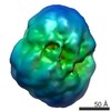 EMDB-7969: |
| Source |
|

