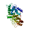
PDB-12qh: Room Temperature X-Ray Structure of SARS-CoV-2 Main Protease in Complex with noncovalent inhibitor KK-1 Method: X-RAY DIFFRACTION / Resolution: 1.8 Å
PDB-12sv: Room Temperature X-Ray Structure of SARS-CoV-2 Main Protease in Complex with noncovalent inhibitor KK-2 Method: X-RAY DIFFRACTION / Resolution: 2 Å
PDB-12ta: Room Temperature X-Ray Structure of SARS-CoV-2 Main Protease in Complex with noncovalent inhibitor KK-3 Method: X-RAY DIFFRACTION / Resolution: 1.8 Å
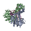
PDB-12wc: Room Temperature X-Ray Structure of SARS-CoV-2 Main Protease in Complex with noncovalent inhibitor KK-7 Method: X-RAY DIFFRACTION / Resolution: 1.8 Å
PDB-12xd: Room Temperature X-Ray Structure of SARS-CoV-2 Main Protease in Complex with noncovalent inhibitor KB-1 Method: X-RAY DIFFRACTION / Resolution: 2 Å
PDB-12yf: Room Temperature X-Ray Structure of SARS-CoV-2 Main Protease in Complex with noncovalent inhibitor KB-2 Method: X-RAY DIFFRACTION / Resolution: 2 Å
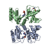
PDB-12zg: Room Temperature X-Ray Structure of SARS-CoV-2 Main Protease in Complex with noncovalent inhibitor KB-3 Method: X-RAY DIFFRACTION / Resolution: 1.8 Å
PDB-12zl: Room Temperature X-Ray Structure of SARS-CoV-2 Main Protease in Complex with noncovalent inhibitor KB-4 Method: X-RAY DIFFRACTION / Resolution: 1.8 Å
PDB-13bw: Room Temperature X-Ray Structure of SARS-CoV-2 Main Protease in Complex with noncovalent inhibitor KB-6 Method: X-RAY DIFFRACTION / Resolution: 1.8 Å
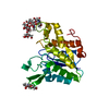
PDB-13cl: Room Temperature X-Ray Structure of SARS-CoV-2 Main Protease in Complex with noncovalent inhibitor KB-0-1 Method: X-RAY DIFFRACTION / Resolution: 2 Å
PDB-13fa: Room Temperature X-Ray Structure of SARS-CoV-2 Main Protease in Complex with noncovalent inhibitor KB-0-2 Method: X-RAY DIFFRACTION / Resolution: 2 Å
PDB-13ho: Joint X-ray/neutron structure of deuterated SARS-CoV-2 main protease (MPro) in complex with noncovalent inhibitor KB-5 Method: X-RAY DIFFRACTION / NEUTRON DIFFRACTION / Resolution: 1.9 Å
Chemicals
PDB-1dcq: CRYSTAL STRUCTURE OF THE ARF-GAP DOMAIN AND ANKYRIN REPEATS OF PAPBETA.
PDB-1dc0: CRYSTAL STRUCTURE OF AN A/B-DNA INTERMEDIATE CATGGGCCCATG
PDB-1dc1: RESTRICTION ENZYME BSOBI/DNA COMPLEX STRUCTURE: ENCIRCLEMENT OF THE DNA AND HISTIDINE-CATALYZED HYDROLYSIS WITHIN A CANONICAL RESTRICTION ENZYME FOLD
PDB-1dc3: STRUCTURAL ANALYSIS OF GLYCERALDEHYDE 3-PHOSPHATE DEHYDROGENASE FROM ESCHERICHIA COLI: DIRECT EVIDENCE FOR SUBSTRATE BINDING AND COFACTOR-INDUCED CONFORMATIONAL CHANGES
PDB-1dek: DEOXYNUCLEOSIDE MONOPHOSPHATE KINASE COMPLEXED WITH DEOXY-GMP
PDB-1dex: RHAMNOGALACTURONAN ACETYLESTERASE FROM ASPERGILLUS ACULEATUS AT 1.9 A RESOLUTION
PDB-1dey: NMR SOLUTION STRUCTURE OF CO(II)-BLEOMYCIN A2
PDB-1de9: HUMAN APE1 ENDONUCLEASE WITH BOUND ABASIC DNA AND MN2+ ION
PDB-1dfa: CRYSTAL STRUCTURE OF PI-SCEI IN C2 SPACE GROUP
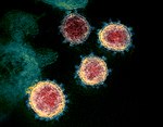
In the structure databanks used in Yorodumi, some data are registered as the other names, "COVID-19 virus" and "2019-nCoV". Here are the details of the virus and the list of structure data.
Jan 31, 2019. EMDB accession codes are about to change! (news from PDBe EMDB page)
EMDB accession codes are about to change! (news from PDBe EMDB page)
The allocation of 4 digits for EMDB accession codes will soon come to an end. Whilst these codes will remain in use, new EMDB accession codes will include an additional digit and will expand incrementally as the available range of codes is exhausted. The current 4-digit format prefixed with “EMD-” (i.e. EMD-XXXX) will advance to a 5-digit format (i.e. EMD-XXXXX), and so on. It is currently estimated that the 4-digit codes will be depleted around Spring 2019, at which point the 5-digit format will come into force.
The EM Navigator/Yorodumi systems omit the EMD- prefix.
Related info.:Q: What is EMD? / ID/Accession-code notation in Yorodumi/EM Navigator
 Movie
Movie Controller
 Structure viewers
 About Yorodumi Papers


 Authors
 External links









 Keywords
 severe acute respiratory syndrome coronavirus 2