 Movie
Movie Controller
 Structure viewers
 About Yorodumi Papers


+Search query
-Structure paper
| Title | Molecular basis of ligand binding and receptor activation at the human A adenosine receptor. |
|---|---|
| Journal, issue, pages | Nat Commun, Vol. 16, Issue 1, Page 7674, Year 2025 |
| Publish date | Aug 18, 2025 |
 Authors Authors | Liudi Zhang / Jesse I Mobbs / Felix M Bennetts / Hariprasad Venugopal / Anh T N Nguyen / Arthur Christopoulos / Daan van der Es / Laura H Heitman / Lauren T May / Alisa Glukhova / David M Thal /   |
| PubMed Abstract | Adenosine receptors (ARs: AAR, AAR, AAR, and AAR) are crucial therapeutic targets; however, developing selective, efficacious drugs for them remains a significant challenge. Here, we present high- ...Adenosine receptors (ARs: AAR, AAR, AAR, and AAR) are crucial therapeutic targets; however, developing selective, efficacious drugs for them remains a significant challenge. Here, we present high-resolution cryo-electron microscopy (cryo-EM) structures of the human AAR in three distinct functional states: bound to the endogenous agonist adenosine, the clinically relevant agonist Piclidenoson, and the covalent antagonist LUF7602. These structures, complemented by mutagenesis and pharmacological studies, reveal an AAR activation mechanism that involves an extensive hydrogen bond network from the extracellular surface down to the orthosteric binding site. In addition, we identify a cryptic pocket that accommodates the N-iodobenzyl group of Piclidenoson through a ligand-dependent conformational change of M174. Our comprehensive structural and functional characterisation of AAR advances our understanding of adenosine receptor pharmacology and establishes a foundation for developing more selective therapeutics for various disorders, including inflammatory diseases, cancer, and glaucoma. |
 External links External links | Nat Commun / PubMed:40825947 / PubMed Central |
| Methods | EM (single particle) |
| Resolution | 2.6 - 3.6 Å |
| Structure data | EMDB-47879, PDB-9ebh: EMDB-47880, PDB-9ebi:  EMDB-47994: Focused refinement map of the human A3 adenosine receptor bound to adenosine (receptor only)  EMDB-47998: Focused refinement map of the human A3 adenosine receptor bound to Piclidenoson (receptor only)  EMDB-48063: Consensus map for A3AR-LUF7602 complex.  EMDB-48064: Focused refinement map of the structure of a human adenosine A3 receptor complex bound to the covalent antagonist LUF7602 (receptor only) EMDB-48065, PDB-9ehs: |
| Chemicals |  ChemComp-ADN:  ChemComp-Q8L: 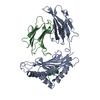 PDB-1bii: |
| Source |
|
 Keywords Keywords | MEMBRANE PROTEIN / G protein-coupled receptor / adenosine binding / seven transmembrane protein / SIGNALING PROTEIN |


 homo sapiens (human)
