ムービー
ムービー コントローラー
 構造ビューア
 万見文献について


+検索条件
-Structure paper
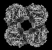
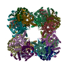
| タイトル | Dimerization of a 5-kDa domain defines the architecture of the 5-MDa gammaproteobacterial pyruvate dehydrogenase complex. |
|---|---|
| ジャーナル・号・ページ | Sci Adv, Vol. 10, Issue 6, Page eadj6358, Year 2024 |
| 掲載日 | 2024年2月9日 |
 著者 著者 | Sarah Meinhold / Rafal Zdanowicz / Christoph Giese / Rudi Glockshuber /  |
| PubMed 要旨 | The pyruvate dehydrogenase complex (PDHc) is a ~5 MDa assembly of the catalytic subunits pyruvate dehydrogenase (E1), dihydrolipoamide acetyltransferase (E2), and dihydrolipoamide dehydrogenase (E3). ...The pyruvate dehydrogenase complex (PDHc) is a ~5 MDa assembly of the catalytic subunits pyruvate dehydrogenase (E1), dihydrolipoamide acetyltransferase (E2), and dihydrolipoamide dehydrogenase (E3). The PDHc core is a cubic complex of eight E2 homotrimers. Homodimers of the peripheral subunits E1 and E3 associate with the core by binding to the peripheral subunit binding domain (PSBD) of E2. Previous reports indicated that 12 E1 dimers and 6 E3 dimers bind to the 24-meric E2 core. Using an assembly arrested E2 homotrimer (E2), we show that two of the three PSBDs in the E2 dimerize, that each PSBD dimer cooperatively binds two E1 dimers, and that E3 dimers only bind to the unpaired PSBD in E2. This mechanism is preserved in wild-type PDHc, with an E1 dimer:E2 monomer:E3 dimer stoichiometry of 16:24:8. The conserved PSBD dimer interface indicates that PSBD dimerization is the previously unrecognized architectural determinant of gammaproteobacterial PDHc megacomplexes. |
 リンク リンク | Sci Adv / PubMed:38324697 / PubMed Central |
| 手法 | EM (単粒子) / X線回折 |
| 解像度 | 1.64 - 3.52 Å |
| 構造データ | EMDB-17119, PDB-8orb:  EMDB-17126: E. coli pyruvate dehydrogenase (E1) in complex with dihydrolipoamide acetyltransferase (E2) peripheral subunit-binding domain. 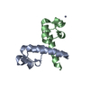 PDB-8oqj: 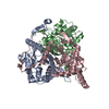 PDB-8osy: |
| 化合物 |  ChemComp-ZN:  ChemComp-HOH: |
| 由来 |
|
 キーワード キーワード | PROTEIN BINDING / Pyruvate dehydrogenase complex / Binding domain / Dimer / PSBD / TRANSFERASE / PDHc / E2 / cryo-EM / Catalytic domain / Trimer |