 Movie
Movie Controller

[English] 日本語
 Yorodumi
- PDB-8orb: 24-meric catalytic domain of dihydrolipoamide acetyltransferase (... -
+ Open data

- Basic information
| Entry | Database: PDB / ID: 8orb | ||||||
|---|---|---|---|---|---|---|---|
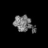
| Title | 24-meric catalytic domain of dihydrolipoamide acetyltransferase (E2) of the E. coli pyruvate dehydrogenase complex. | ||||||
 Components Components | Dihydrolipoyllysine-residue acetyltransferase component of pyruvate dehydrogenase complex | ||||||
 Keywords Keywords | TRANSFERASE / pyruvate dehydrogenase complex / PDHc / E2 / cryo-EM | ||||||
| Function / homology |  Function and homology information Function and homology informationstress response to acid chemical / dihydrolipoyllysine-residue acetyltransferase / dihydrolipoyllysine-residue acetyltransferase activity / pyruvate decarboxylation to acetyl-CoA / lipoic acid binding / pyruvate catabolic process / pyruvate dehydrogenase complex / cytoplasm Similarity search - Function | ||||||
| Biological species |  | ||||||
| Method | ELECTRON MICROSCOPY / single particle reconstruction / cryo EM / Resolution: 3.25 Å | ||||||
 Authors Authors | Zdanowicz, R. / Meinhold, S. / Glockshuber, R. | ||||||
| Funding support |  Switzerland, 1items Switzerland, 1items
| ||||||
 Citation Citation | Journal: Sci Adv / Year: 2024 Title: Dimerization of a 5-kDa domain defines the architecture of the 5-MDa gammaproteobacterial pyruvate dehydrogenase complex. Authors: Sarah Meinhold / Rafal Zdanowicz / Christoph Giese / Rudi Glockshuber / Abstract: The pyruvate dehydrogenase complex (PDHc) is a ~5 MDa assembly of the catalytic subunits pyruvate dehydrogenase (E1), dihydrolipoamide acetyltransferase (E2), and dihydrolipoamide dehydrogenase (E3). ...The pyruvate dehydrogenase complex (PDHc) is a ~5 MDa assembly of the catalytic subunits pyruvate dehydrogenase (E1), dihydrolipoamide acetyltransferase (E2), and dihydrolipoamide dehydrogenase (E3). The PDHc core is a cubic complex of eight E2 homotrimers. Homodimers of the peripheral subunits E1 and E3 associate with the core by binding to the peripheral subunit binding domain (PSBD) of E2. Previous reports indicated that 12 E1 dimers and 6 E3 dimers bind to the 24-meric E2 core. Using an assembly arrested E2 homotrimer (E2), we show that two of the three PSBDs in the E2 dimerize, that each PSBD dimer cooperatively binds two E1 dimers, and that E3 dimers only bind to the unpaired PSBD in E2. This mechanism is preserved in wild-type PDHc, with an E1 dimer:E2 monomer:E3 dimer stoichiometry of 16:24:8. The conserved PSBD dimer interface indicates that PSBD dimerization is the previously unrecognized architectural determinant of gammaproteobacterial PDHc megacomplexes. | ||||||
| History |
|
- Structure visualization
| Structure viewer | Molecule: MolmilJmol/JSmol |
|---|
- Downloads & links
-Download
| PDBx/mmCIF format | 8orb.cif.gz | 1 MB | Display | PDBx/mmCIF format |
|---|---|---|---|---|
| PDB format | pdb8orb.ent.gz | 908.5 KB | Display | PDB format |
| PDBx/mmJSON format | 8orb.json.gz | Tree view | PDBx/mmJSON format | |
| Others |  Other downloads Other downloads |
-Validation report
| Arichive directory | https://data.pdbj.org/pub/pdb/validation_reports/or/8orbftp://data.pdbj.org/pub/pdb/validation_reports/or/8orb | HTTPS FTP |
|---|
-Related structure data
| Related structure data |  17119MC 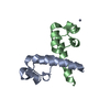 8oqjC 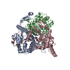 8osyC M: map data used to model this data C: citing same article ( |
|---|---|
| Similar structure data |
-Links
 PDBj
PDBj

- Assembly
| Deposited unit | 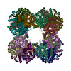
|
|---|---|
| 1 |
|
-Components
| #1: Protein | Mass: 27491.049 Da / Num. of mol.: 24 Source method: isolated from a genetically manipulated source Source: (gene. exp.) References: UniProt: P06959, dihydrolipoyllysine-residue acetyltransferase |
|---|
-Experimental details
-Experiment
| Experiment | Method: ELECTRON MICROSCOPY |
|---|---|
| EM experiment | Aggregation state: PARTICLE / 3D reconstruction method: single particle reconstruction |
- Sample preparation
| Component | Name: 24-meric catalytic domain of dihydrolipoamide acetyltransferase (E2) of the E. coli pyruvate dehydrogenase complex Type: COMPLEX Details: Wild-type E2 protein was truncated to contain only the catalytic domain Entity ID: all / Source: RECOMBINANT |
|---|---|
| Molecular weight | Value: 0.66 MDa / Experimental value: NO |
| Source (natural) | Organism: |
| Source (recombinant) | Organism: |
| Buffer solution | pH: 7.4 |
| Specimen | Embedding applied: NO / Shadowing applied: NO / Staining applied: NO / Vitrification applied: YES |
| Specimen support | Grid material: COPPER / Grid mesh size: 300 divisions/in. / Grid type: Quantifoil R2/2 |
| Vitrification | Instrument: FEI VITROBOT MARK IV / Cryogen name: ETHANE / Humidity: 100 % / Chamber temperature: 277 K |
- Electron microscopy imaging
| Experimental equipment |  Model: Titan Krios / Image courtesy: FEI Company |
|---|---|
| Microscopy | Model: FEI TITAN KRIOS |
| Electron gun | Electron source:  FIELD EMISSION GUN / Accelerating voltage: 300 kV / Illumination mode: FLOOD BEAM FIELD EMISSION GUN / Accelerating voltage: 300 kV / Illumination mode: FLOOD BEAM |
| Electron lens | Mode: BRIGHT FIELD / Nominal defocus max: 2800 nm / Nominal defocus min: 1400 nm |
| Image recording | Electron dose: 80 e/Å2 / Detector mode: COUNTING / Film or detector model: GATAN K2 SUMMIT (4k x 4k) |
- Processing
| EM software |
| |||||||||||||||||||||||||
|---|---|---|---|---|---|---|---|---|---|---|---|---|---|---|---|---|---|---|---|---|---|---|---|---|---|---|
| CTF correction | Type: PHASE FLIPPING AND AMPLITUDE CORRECTION | |||||||||||||||||||||||||
| Symmetry | Point symmetry: O (octahedral) | |||||||||||||||||||||||||
| 3D reconstruction | Resolution: 3.25 Å / Resolution method: FSC 0.143 CUT-OFF / Num. of particles: 278268 / Symmetry type: POINT |
