 Movie
Movie Controller
 Structure viewers
 About Yorodumi Papers


+Search query
-Structure paper
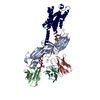
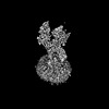
| Title | Tail engagement of arrestin at the glucagon receptor. |
|---|---|
| Journal, issue, pages | Nature, Vol. 620, Issue 7975, Page 904-910, Year 2023 |
| Publish date | Aug 9, 2023 |
 Authors Authors | Kun Chen / Chenhui Zhang / Shuling Lin / Xinyu Yan / Heng Cai / Cuiying Yi / Limin Ma / Xiaojing Chu / Yuchen Liu / Ya Zhu / Shuo Han / Qiang Zhao / Beili Wu /  |
| PubMed Abstract | Arrestins have pivotal roles in regulating G protein-coupled receptor (GPCR) signalling by desensitizing G protein activation and mediating receptor internalization. It has been proposed that the ...Arrestins have pivotal roles in regulating G protein-coupled receptor (GPCR) signalling by desensitizing G protein activation and mediating receptor internalization. It has been proposed that the arrestin binds to the receptor in two different conformations, 'tail' and 'core', which were suggested to govern distinct processes of receptor signalling and trafficking. However, little structural information is available for the tail engagement of the arrestins. Here we report two structures of the glucagon receptor (GCGR) bound to β-arrestin 1 (βarr1) in glucagon-bound and ligand-free states. These structures reveal a receptor tail-engaged binding mode of βarr1 with many unique features, to our knowledge, not previously observed. Helix VIII, instead of the receptor core, has a major role in accommodating βarr1 by forming extensive interactions with the central crest of βarr1. The tail-binding pose is further defined by a close proximity between the βarr1 C-edge and the receptor helical bundle, and stabilized by a phosphoinositide derivative that bridges βarr1 with helices I and VIII of GCGR. Lacking any contact with the arrestin, the receptor core is in an inactive state and loosely binds to glucagon. Further functional studies suggest that the tail conformation of GCGR-βarr governs βarr recruitment at the plasma membrane and endocytosis of GCGR, and provides a molecular basis for the receptor forming a super-complex simultaneously with G protein and βarr to promote sustained signalling within endosomes. These findings extend our knowledge about the arrestin-mediated modulation of GPCR functionalities. |
 External links External links | Nature / PubMed:37558880 / PubMed Central |
| Methods | EM (single particle) |
| Resolution | 3.3 - 3.5 Å |
| Structure data | EMDB-36606, PDB-8jru: EMDB-36607, PDB-8jrv: |
| Chemicals |  ChemComp-PIO: |
| Source |
|
 Keywords Keywords | MEMBRANE PROTEIN / Complex structure / glucagon receptor / beta-arrestin 1 / ligand-free / glucagon |


 homo sapiens (human)
 escherichia phage ecszw-2 (virus)