 Movie
Movie Controller
 Structure viewers
 About Yorodumi Papers


+Search query
-Structure paper
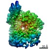
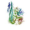
| Title | Structural Analysis of Botulinum Neurotoxins Type B and E by Cryo-EM. |
|---|---|
| Journal, issue, pages | Toxins (Basel), Vol. 14, Issue 1, Year 2021 |
| Publish date | Dec 23, 2021 |
 Authors Authors | Sara Košenina / Markel Martínez-Carranza / Jonathan R Davies / Geoffrey Masuyer / Pål Stenmark /   |
| PubMed Abstract | Botulinum neurotoxins (BoNTs) are the causative agents of a potentially lethal paralytic disease targeting cholinergic nerve terminals. Multiple BoNT serotypes exist, with types A, B and E being the ...Botulinum neurotoxins (BoNTs) are the causative agents of a potentially lethal paralytic disease targeting cholinergic nerve terminals. Multiple BoNT serotypes exist, with types A, B and E being the main cause of human botulism. Their extreme toxicity has been exploited for cosmetic and therapeutic uses to treat a wide range of neuromuscular disorders. Although naturally occurring BoNT types share a common end effect, their activity varies significantly based on the neuronal cell-surface receptors and intracellular SNARE substrates they target. These properties are the result of structural variations that have traditionally been studied using biophysical methods such as X-ray crystallography. Here, we determined the first structures of botulinum neurotoxins using single-particle cryogenic electron microscopy. The maps obtained at 3.6 and 3.7 Å for BoNT/B and /E, respectively, highlight the subtle structural dynamism between domains, and of the binding domain in particular. This study demonstrates how the recent advances made in the field of single-particle electron microscopy can be applied to bacterial toxins of clinical relevance and the botulinum neurotoxin family in particular. |
 External links External links | Toxins (Basel) / PubMed:35050991 / PubMed Central |
| Methods | EM (single particle) |
| Resolution | 3.6 - 3.7 Å |
| Structure data | EMDB-13946, PDB-7qfp: EMDB-13947, PDB-7qfq: |
| Source |
|
 Keywords Keywords | TOXIN / clostridium botulinum / neurotoxin |


 clostridium botulinum (bacteria)