 ムービー
ムービー コントローラー
 構造ビューア
 万見文献について


+検索条件
-Structure paper
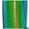
| タイトル | The α-synuclein hereditary mutation E46K unlocks a more stable, pathogenic fibril structure. |
|---|---|
| ジャーナル・号・ページ | Proc Natl Acad Sci U S A, Vol. 117, Issue 7, Page 3592-3602, Year 2020 |
| 掲載日 | 2020年2月18日 |
 著者 著者 | David R Boyer / Binsen Li / Chuanqi Sun / Weijia Fan / Kang Zhou / Michael P Hughes / Michael R Sawaya / Lin Jiang / David S Eisenberg /  |
| PubMed 要旨 | Aggregation of α-synuclein is a defining molecular feature of Parkinson's disease, Lewy body dementia, and multiple systems atrophy. Hereditary mutations in α-synuclein are linked to both ...Aggregation of α-synuclein is a defining molecular feature of Parkinson's disease, Lewy body dementia, and multiple systems atrophy. Hereditary mutations in α-synuclein are linked to both Parkinson's disease and Lewy body dementia; in particular, patients bearing the E46K disease mutation manifest a clinical picture of parkinsonism and Lewy body dementia, and E46K creates more pathogenic fibrils in vitro. Understanding the effect of these hereditary mutations on α-synuclein fibril structure is fundamental to α-synuclein biology. We therefore determined the cryo-electron microscopy (cryo-EM) structure of α-synuclein fibrils containing the hereditary E46K mutation. The 2.5-Å structure reveals a symmetric double protofilament in which the molecules adopt a vastly rearranged, lower energy fold compared to wild-type fibrils. We propose that the E46K misfolding pathway avoids electrostatic repulsion between K46 and K80, a residue pair which form the E46-K80 salt bridge in the wild-type fibril structure. We hypothesize that, under our conditions, the wild-type fold does not reach this deeper energy well of the E46K fold because the E46-K80 salt bridge diverts α-synuclein into a kinetic trap-a shallower, more accessible energy minimum. The E46K mutation apparently unlocks a more stable and pathogenic fibril structure. |
 リンク リンク | Proc Natl Acad Sci U S A / PubMed:32015135 / PubMed Central |
| 手法 | EM (らせん対称) |
| 解像度 | 2.5 Å |
| 構造データ | EMDB-20759, PDB-6ufr: |
| 化合物 |  ChemComp-HOH: |
| 由来 |
|
 キーワード キーワード | PROTEIN FIBRIL / alpha-synuclein / amyloid / fibril / E46K / hereditary mutations / parkinson's disease / lewy body dementia |

 homo sapiens (ヒト)