PDB-6gi0: Crystal structure of the ferric enterobactin esterase (PfeE) from Pseudomonas aeruginosa 手法: X-RAY DIFFRACTION / 解像度: 2 Å
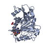
PDB-6gi1: Crystal structure of the ferric enterobactin esterase (pfeE) mutant(S157A) from Pseudomonas aeruginosa in presence of enterobactin 手法: X-RAY DIFFRACTION / 解像度: 1.66 Å
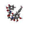
PDB-6gi2: Crystal structure of the ferric enterobactin esterase (pfeE) mutant(S157A) from Pseudomonas aeruginosa in complex with Tris-catechol vector 手法: X-RAY DIFFRACTION / 解像度: 1.49 Å
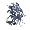
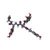
PDB-6gi5: Crystal structure of the ferric enterobactin esterase (PfeE) from Pseudomonas aeruginosa in complex with the tris-catechol vector 手法: X-RAY DIFFRACTION / 解像度: 3.11 Å
 ムービー
ムービー コントローラー
 構造ビューア
 万見文献について


 著者
著者 リンク
リンク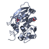


 キーワード
キーワード
 pseudomonas aeruginosa (緑膿菌)