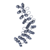
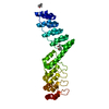
PDB-5le2: Crystal structure of DARPin-DARPin rigid fusion, variant DDD_D12_15_D12_15_D12 Method: X-RAY DIFFRACTION / Resolution: 2.4 Å
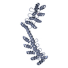
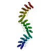
PDB-5le3: Crystal structure of DARPin-DARPin rigid fusion, variant DD_D12_09_D12 Method: X-RAY DIFFRACTION / Resolution: 3.5 Å
PDB-5le4: Crystal structure of DARPin-DARPin rigid fusion, variant DD_D12_11_D12 Method: X-RAY DIFFRACTION / Resolution: 2.35 Å
PDB-5le6: Crystal structure of DARPin-DARPin rigid fusion, variant DD_D12_09_D12 Method: X-RAY DIFFRACTION / Resolution: 1.8 Å
PDB-5le7: Crystal structure of DARPin-DARPin rigid fusion, variant DD_D12_13_D12 Method: X-RAY DIFFRACTION / Resolution: 2.104 Å
PDB-5le8: Crystal structure of DARPin-DARPin rigid fusion, variant DD_D12_15_D12 Method: X-RAY DIFFRACTION / Resolution: 1.78 Å
PDB-5le9: Crystal structure of DARPin-DARPin rigid fusion, variant DD_Off7_09_3G124 Method: X-RAY DIFFRACTION / Resolution: 1.85 Å
PDB-5lea: Crystal structure of DARPin-DARPin rigid fusion, variant DD_Off7_12_3G124 Method: X-RAY DIFFRACTION / Resolution: 2.4 Å
PDB-5leb: Crystal structure of DARPin-DARPin rigid fusion, variant DDD_D12_06_D12_06_D12 Method: X-RAY DIFFRACTION / Resolution: 2.3 Å
PDB-5lec: Crystal structure of DARPin-DARPin rigid fusion, variant DDD_D12_12_D12_12_D12 Method: X-RAY DIFFRACTION / Resolution: 2.506 Å
PDB-5led: Crystal structure of DARPin-DARPin rigid fusion, variant DDD_D12_12_D12_12_D12 Method: X-RAY DIFFRACTION / Resolution: 2.6 Å
PDB-5lee: Crystal structure of DARPin-DARPin rigid fusion, variant DDD_D12_12_D12_12_D12 Method: X-RAY DIFFRACTION / Resolution: 2.401 Å
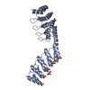
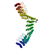
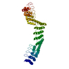
PDB-5lel: Crystal structure of DARPin-DARPin rigid fusion, variant DD_Off7_10_3G124 in complex with Maltose-binding Protein and Green Fluorescent Protein Method: X-RAY DIFFRACTION / Resolution: 3.1 Å
PDB-5lem: Crystal structure of DARPin-DARPin rigid fusion, variant DD_Off7_11_3G124 in complex with Maltose-binding Protein and Green Fluorescent Protein Method: X-RAY DIFFRACTION / Resolution: 2.98 Å
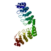
PDB-5lw2: Crystal structure of DARPin 5m3_D12 Method: X-RAY DIFFRACTION / Resolution: 1.75 Å
DE NOVO PROTEIN / X-ray crystallography; designed ankyrin repeat proteins; protein design; protein engineering; rigid domain fusions / designed ankyrin repeat proteins / protein design / protein engineering / rigid domain fusions / designed ankyrin repeat proteins; protein design; protein engineering; rigid domain fusions / FLUORESCENT PROTEIN
+
About Yorodumi Papers
-
News
-
Feb 9, 2022. New format data for meta-information of EMDB entries
New format data for meta-information of EMDB entries
Version 3 of the EMDB header file is now the official format.
The previous official version 1.9 will be removed from the archive.
In the structure databanks used in Yorodumi, some data are registered as the other names, "COVID-19 virus" and "2019-nCoV". Here are the details of the virus and the list of structure data.
Jan 31, 2019. EMDB accession codes are about to change! (news from PDBe EMDB page)
EMDB accession codes are about to change! (news from PDBe EMDB page)
The allocation of 4 digits for EMDB accession codes will soon come to an end. Whilst these codes will remain in use, new EMDB accession codes will include an additional digit and will expand incrementally as the available range of codes is exhausted. The current 4-digit format prefixed with “EMD-” (i.e. EMD-XXXX) will advance to a 5-digit format (i.e. EMD-XXXXX), and so on. It is currently estimated that the 4-digit codes will be depleted around Spring 2019, at which point the 5-digit format will come into force.
The EM Navigator/Yorodumi systems omit the EMD- prefix.
Related info.:Q: What is EMD? / ID/Accession-code notation in Yorodumi/EM Navigator
 Movie
Movie Controller
 Structure viewers
 About Yorodumi Papers


 Authors
 External links


 Keywords

 aequorea victoria (jellyfish)