 Movie
Movie Controller
 Structure viewers
 About Yorodumi Papers


+Search query
-Structure paper
| Title | Crystal structure of the PEG-bound SH3 domain of myosin IB from Entamoeba histolytica reveals its mode of ligand recognition |
|---|---|
| Journal, issue, pages | Acta Crystallogr D Struct Biol, Vol. 73, Page 672-682, Year 2017 |
| Publish date | Apr 13, 2017 (structure data deposition date) |
 Authors Authors | Gautam, G. / Rehman, S.A.A. / Pandey, P. / Gourinath, S. |
 External links External links | Acta Crystallogr D Struct Biol / PubMed:28777082 |
| Methods | X-ray diffraction |
| Resolution | 1.72 - 1.78 Å |
| Structure data |  PDB-5xg9:  PDB-5xgg: |
| Chemicals |  ChemComp-1PE:  ChemComp-PEU: 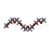 ChemComp-PG6:  ChemComp-SO4:  ChemComp-HOH: |
| Source |
|
 Keywords Keywords | CONTRACTILE PROTEIN / SH3 / MyosinI / Entamoeba histolytica / PEG-bound SH3 complex / EhMySH3 |

 entamoeba histolytica (eukaryote)