Movie
Movie Controller
 Structure viewers
 About Yorodumi Papers


+Search query
-Structure paper
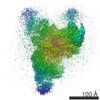
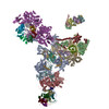
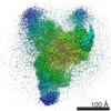
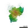
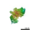
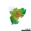
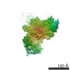
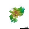
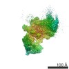
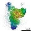
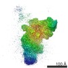
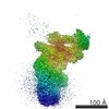
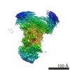
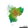
| Title | The 3.8 Å structure of the U4/U6.U5 tri-snRNP: Insights into spliceosome assembly and catalysis. |
|---|---|
| Journal, issue, pages | Science, Vol. 351, Issue 6272, Page 466-475, Year 2016 |
| Publish date | Jan 29, 2016 |
 Authors Authors | Ruixue Wan / Chuangye Yan / Rui Bai / Lin Wang / Min Huang / Catherine C L Wong / Yigong Shi /  |
| PubMed Abstract | Splicing of precursor messenger RNA is accomplished by a dynamic megacomplex known as the spliceosome. Assembly of a functional spliceosome requires a preassembled U4/U6.U5 tri-snRNP complex, which ...Splicing of precursor messenger RNA is accomplished by a dynamic megacomplex known as the spliceosome. Assembly of a functional spliceosome requires a preassembled U4/U6.U5 tri-snRNP complex, which comprises the U5 small nuclear ribonucleoprotein (snRNP), the U4 and U6 small nuclear RNA (snRNA) duplex, and a number of protein factors. Here we report the three-dimensional structure of a Saccharomyces cerevisiae U4/U6.U5 tri-snRNP at an overall resolution of 3.8 angstroms by single-particle electron cryomicroscopy. The local resolution for the core regions of the tri-snRNP reaches 3.0 to 3.5 angstroms, allowing construction of a refined atomic model. Our structure contains U5 snRNA, the extensively base-paired U4/U6 snRNA, and 30 proteins including Prp8 and Snu114, which amount to 8495 amino acids and 263 nucleotides with a combined molecular mass of ~1 megadalton. The catalytic nucleotide U80 from U6 snRNA exists in an inactive conformation, stabilized by its base-pairing interactions with U4 snRNA and protected by Prp3. Pre-messenger RNA is bound in the tri-snRNP through base-pairing interactions with U6 snRNA and loop I of U5 snRNA. This structure, together with that of the spliceosome, reveals the molecular choreography of the snRNAs in the activation process of the spliceosomal ribozyme. |
 External links External links | Science / PubMed:26743623 |
| Methods | EM (single particle) |
| Resolution | 3.46 - 7.9 Å |
| Structure data | EMDB-6561: The cryo-EM structure of yeast spliceosomal U4/U6.U5 tri-snRNP at 3.8 A EMDB-6562: The cryo-EM structure of yeast spliceosomal U4/U6.U5 tri-snRNP (improved map for U4/U6 and U5 RNA region at 3.75 A) EMDB-6563: The cryo-EM structure of yeast spliceosomal U4/U6.U5 tri-snRNP (improved map for U4/U6 RNA region at 3.57 A) EMDB-6564: The cryo-EM structure of yeast spliceosomal U4/U6.U5 tri-snRNP (improved map for Prp31 region at 3.52 A) EMDB-6565: The cryo-EM structure of yeast spliceosomal U4/U6.U5 tri-snRNP (improved map for Prp8 region at 3.49 A) EMDB-6566: The cryo-EM structure of yeast spliceosomal U4/U6.U5 tri-snRNP (improved map for Prp3 region at 3.46 A) EMDB-6567: The cryo-EM structure of yeast spliceosomal U4/U6.U5 tri-snRNP (improved map for Prp4 region at 3.61 A) EMDB-6568: The cryo-EM structure of yeast spliceosomal U4/U6.U5 tri-snRNP (improved map for Prp4 WD40 region at 3.70 A) EMDB-6569: The cryo-EM structure of yeast spliceosomal U4/U6.U5 tri-snRNP (improved map for U5 RNA region at 3.70 A) EMDB-6570: The cryo-EM structure of yeast spliceosomal U4/U6.U5 tri-snRNP (improved map for U5 Snu114 region at 3.77 A) EMDB-6571: The cryo-EM structure of yeast spliceosomal U4/U6.U5 tri-snRNP (improved map for U5 Sm ring region at 4.7 A) |
| Chemicals |  ChemComp-GTP:  ChemComp-M7M: |
| Source |
|
 Keywords Keywords | TRANSCRIPTION / U4/U6.U5 tri-snRNP / pre-mRNA |