 ムービー
ムービー コントローラー
 構造ビューア
 万見文献について


+検索条件
-Structure paper
| タイトル | CTER-rapid estimation of CTF parameters with error assessment. |
|---|---|
| ジャーナル・号・ページ | Ultramicroscopy, Vol. 140, Page 9-19, Year 2014 |
| 掲載日 | 2014年2月7日 |
 著者 著者 | Pawel A Penczek / Jia Fang / Xueming Li / Yifan Cheng / Justus Loerke / Christian M T Spahn /   |
| PubMed 要旨 | In structural electron microscopy, the accurate estimation of the Contrast Transfer Function (CTF) parameters, particularly defocus and astigmatism, is of utmost importance for both initial ...In structural electron microscopy, the accurate estimation of the Contrast Transfer Function (CTF) parameters, particularly defocus and astigmatism, is of utmost importance for both initial evaluation of micrograph quality and for subsequent structure determination. Due to increases in the rate of data collection on modern microscopes equipped with new generation cameras, it is also important that the CTF estimation can be done rapidly and with minimal user intervention. Finally, in order to minimize the necessity for manual screening of the micrographs by a user it is necessary to provide an assessment of the errors of fitted parameters values. In this work we introduce CTER, a CTF parameters estimation method distinguished by its computational efficiency. The efficiency of the method makes it suitable for high-throughput EM data collection, and enables the use of a statistical resampling technique, bootstrap, that yields standard deviations of estimated defocus and astigmatism amplitude and angle, thus facilitating the automation of the process of screening out inferior micrograph data. Furthermore, CTER also outputs the spatial frequency limit imposed by reciprocal space aliasing of the discrete form of the CTF and the finite window size. We demonstrate the efficiency and accuracy of CTER using a data set collected on a 300kV Tecnai Polara (FEI) using the K2 Summit DED camera in super-resolution counting mode. Using CTER we obtained a structure of the 80S ribosome whose large subunit had a resolution of 4.03Å without, and 3.85Å with, inclusion of astigmatism parameters. |
 リンク リンク | Ultramicroscopy / PubMed:24562077 / PubMed Central |
| 手法 | EM (単粒子) |
| 解像度 | 3.85 Å |
| 構造データ | 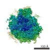 EMDB-5889: |
| 由来 |
|
 Homo sapiens (ヒト)