 Movie
Movie Controller
 Structure viewers
 About Yorodumi Papers


+Search query
-Structure paper
| Title | An externalized polypeptide partitions between two distinct sites on genome-released poliovirus particles. |
|---|---|
| Journal, issue, pages | J Virol, Vol. 85, Issue 19, Page 9974-9983, Year 2011 |
| Publish date | Jul 20, 2011 |
 Authors Authors | Jun Lin / Naiqian Cheng / Marie Chow / David J Filman / Alasdair C Steven / James M Hogle / David M Belnap /  |
| PubMed Abstract | During cell entry, native poliovirus (160S) converts to a cell-entry intermediate (135S) particle, resulting in the externalization of capsid proteins VP4 and the amino terminus of VP1 (residues 1 to ...During cell entry, native poliovirus (160S) converts to a cell-entry intermediate (135S) particle, resulting in the externalization of capsid proteins VP4 and the amino terminus of VP1 (residues 1 to 53). Externalization of these entities is followed by release of the RNA genome (uncoating), leaving an empty (80S) particle. The antigen-binding fragment (Fab) of a monospecific peptide 1 (P1) antibody, which was raised against a peptide corresponding to amino-terminal residues 24 to 40 of VP1, was utilized to track the location of the amino terminus of VP1 in the 135S and 80S states of poliovirus particles via cryogenic electron microscopy (cryo-EM) and three-dimensional image reconstruction. On 135S, P1 Fabs bind to a prominent feature on the external surface known as the "propeller tip." In contrast, our initial 80S-P1 reconstruction showed P1 Fabs also binding to a second site, at least 50 Å distant, at the icosahedral 2-fold axes. Further analysis showed that the overall population of 80S-P1 particles consisted of three kinds of capsids: those with P1 Fabs bound only at the propeller tips, P1 Fabs bound only at the 2-fold axes, or P1 Fabs simultaneously bound at both positions. Our results indicate that, in 80S particles, a significant fraction of VP1 can deviate from icosahedral symmetry. Hence, this portion of VP1 does not change conformation synchronously when switching from the 135S state. These conclusions are compatible with previous observations of multiple conformations of the 80S state and suggest that movement of the amino terminus of VP1 has a role in uncoating. Similar deviations from icosahedral symmetry may be biologically significant during other viral transitions. |
 External links External links | J Virol / PubMed:21775460 / PubMed Central |
| Methods | EM (single particle) |
| Resolution | 12.0 - 26.0 Å |
| Structure data | 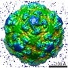 EMDB-5280: 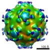 EMDB-5282: 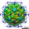 EMDB-5283: 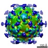 EMDB-5284: 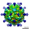 EMDB-5285: 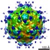 EMDB-5286: |
